Antigen NY-CO-13, BCC7, Cellular Tumor Antigen p53, LFS1, TP53, Transformation Related Protein 53 (TRP53)
The specificity of this monoclonal antibody to its intended target was validated by HuProtTM Array, containing more than 19,000, full-length human proteins. Recognizes a 53kDa protein, which is identified as p53 suppressor gene product. It reacts with the mutant as well as the wild form of p53 protein. p53 is a tumor suppressor gene expressed in a wide variety of tissue types and is involved in regulating cell growth, replication, and apoptosis. It binds to MDM2, SV40 T antigen and human papilloma virus E6 protein. Positive nuclear staining with p53 antibody has been reported to be a negative prognostic factor in breast carcinoma, lung carcinoma, colorectal, and urothelial carcinoma. Anti-p53 positivity has also been used to differentiate uterine serous carcinoma from endometrioid carcinoma as well as to detect intratubular germ cell neoplasia. Mutations involving p53 are found in a wide variety of malignant tumors, including breast, ovarian, bladder, colon, lung, and melanoma.
200ug/ml of Ab purified from Bioreactor Concentrate by Protein A/G. Prepared in 10mM PBS with 0.05% BSA & 0.05% azide. Also available WITHOUT BSA & azide at 1.0mg/ml.
Antibody Type:
Recombinant Monoclonal Antibody
Application Notes:
Immunohistochemistry (Formalin-fixed) (1-2ug/ml for 30 minutes at RT),(Staining of formalin-fixed tissues requires heating tissue sections in 10mM Tris with 1mM EDTA, pH 9.0, for 45 min at 95°C followed by cooling at RT for 20 minutes),Optimal dilutio
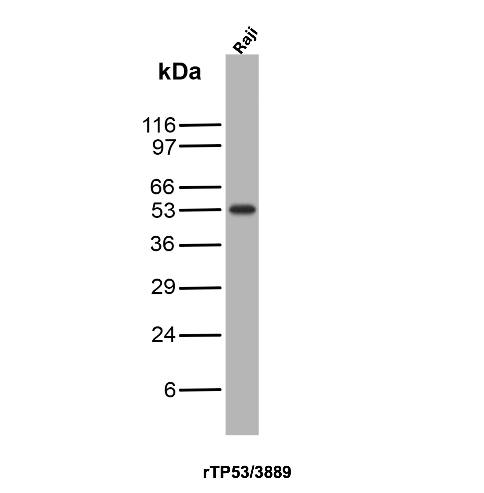
Western Blot Analysis of Raji cell lysate using p53 Recombinant Mouse Monoclonal Antibody (rTP53/3889).
Formalin-fixed, paraffin-embedded human bladder stained with p53 Recombinant Mouse Monoclonal Antibody (rTP53/3889).
SDS-PAGE Analysis of Purified p53 Recombinant Mouse Monoclonal Antibody (rTP53/3889). Confirmation of Purity and Integrity of Antibody.
* VAT and and shipping costs not included. Errors and price changes excepted