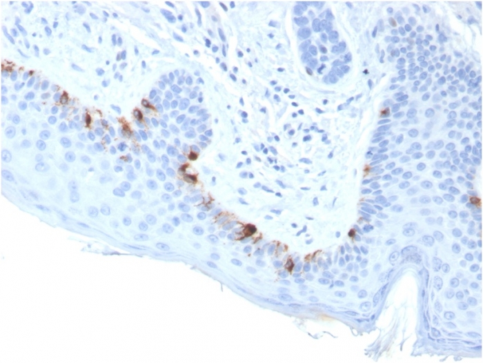
It reacts with a 75kDa melanocyte-specific gene product, identified as Tyrosinase-related protein-1 (TRP-1). It is involved in melanin synthesis. TRP1 is present on the melanosomal membranes of melanoma, normal melanocytes and nevi.Recent evidence suggests that TRP-1 is involved in maintaining stability of tyrosinase protein and modulating its catalytic activity. TRP-1 is also involved in maintenance of melanosome ultrastructure and affects melanocyte proliferation and cell death.
200ug/ml of Ab Purified from Bioreactor Concentrate by Protein A/G. Prepared in 10mM PBS with 0.05% BSA & 0.05% azide. Also available WITHOUT BSA & azide at 1.0mg/ml.
Formalin-fixed, paraffin-embedded human Skin stained with TYRP1-Monospecific Mouse Monoclonal Antibody (TYRP1/3283).
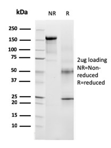
SDS-PAGE Analysis of Purified TYRP1-Monospecific Mouse Monoclonal Antibody (TYRP1/3283). Confirmation of Purity and Integrity of Antibody.
Analysis of Protein Array containing more than 19,000 full-length human proteins using TYRP1-Monospecific Mouse Monoclonal Antibody (TYRP1/3283) Z- and S- Score: The Z-score represents the strength of a signal that a monoclonal antibody (Monoclonal Antibody) (in combination with a fluorescently-tagged anti-IgG secondary antibody) produces when binding to a particular protein on the HuProtTM array. Z-scores are described in units of standard deviations (SD,Aövivs) above the mean value of all signals generated on that array. If targets on HuProtTM are arranged in descending order of the Z-score, the S-score is the difference (also in units of SD,Aövivs) between the Z-score. S-score therefore represents the relative target specificity of a Monoclonal Antibody to its intended target. A Monoclonal Antibody is considered to specific to its intended target, if the Monoclonal Antibody has an S-score of at least 2.5. For example, if a Monoclonal Antibody binds to protein X with a Z-score of 43 and to protein Y with a Z-score of 14, then the S-score for the binding of that Monoclonal Antibody to protein X is equal to 29.
* VAT and and shipping costs not included. Errors and price changes excepted