Amplified in squamous cell carcinoma (AIS), Chronic ulcerative stomatitis protein (CUSP), EEC3, Keratinocyte transcription factor KET, LMS, NBP, p40, P51/P63, p53 like transcription factor, p53-related protein p63, RHS, SHFM4, TAp63alpha, TP53CP, TP53L, TP63, TP73, TP73L, Transformation-related protein 63, Trp53rp1, Trp6,3, Tumor protein 63, Tumor protein p53-like, tumor protein p73-like
p63 is a homolog of the tumor suppressor p53. It is identified in basal cells in the epithelial layers of a variety of tissues, including epidermis, cervix, urothelium, breast and prostate. p63 was detected in nuclei of the basal epithelium in normal prostate glands, however, it was not expressed in malignant tumors of the prostate. As a result, p63 has been reported as a useful marker for differentiating benign from malignant lesions in the prostate, particularly when used in combination with markers of high molecular weight cytokeratins and the prostate-specific marker AMACR (P504S). p63 has also been shown to be a sensitive marker for lung squamous cell carcinomas (SqCC), with a sensitivity of ~90%. Specificity for lung SqCC, vs. lung adenocarcinoma (LADC), is approximately 80%. In breast tissue, p63 has been identified in myoepithelial cells of normal ducts.
200ug/ml of Ab Purified from Bioreactor Concentrate by Protein A/G. Prepared in 10mM PBS with 0.05% BSA & 0.05% azide. Also available WITHOUT BSA & azide at 1.0mg/ml.
Antibody Type:
Monoclonal Antibody
Application Notes:
Immunohistochemistry (Formalin-fixed) (1-2ug/ml for 30 minutes at RT),(Staining of formalin-fixed tissues requires heating tissue sections in 10mM Tris with 1mM EDTA, pH 9.0, for 45 min at 95C followed by cooling at RT for 20 minutes), Optimal dilution
Analysis of Prot
Formalin-fixed, paraffin-embedded human Prostate Carcinoma stained with p63 Mouse Monoclonal Antibody (TP63/2427).
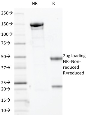
SDS-PAGE Analysis of Purified p63 Mouse Monoclonal Antibody (TP63/2427). Confirmation of Integrity and Purity of Antibody.
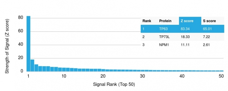
Analysis of Protein Array containing more than 19,000 full-length human proteins using p63 Mouse Monoclonal Antibody (TP63/2427). Z- and S- Score: The Z-score represents the strength of a signal that a monoclonal antibody (Monoclonal Antibody) (in combination with a fluorescently-tagged anti-IgG secondary antibody) produces when binding to a particular protein on the HuProtTM array. Z-scores are described in units of standard deviations (SD,Aövivs) above the mean value of all signals generated on that array. If targets on HuProtTM are arranged in descending order of the Z-score, the S-score is the difference (also in units of SD,Aövivs) between the Z-score. S-score therefore represents the relative target specificity of a Monoclonal Antibody to its intended target. A Monoclonal Antibody is considered to specific to its intended target, if the Monoclonal Antibody has an S-score of at least 2.5. For example, if a Monoclonal Antibody binds to protein X with a Z-score of 43 and to protein Y with a Z-score of 14, then the S-score for the binding of that Monoclonal Antibody to protein X is equal to 29.
* VAT and and shipping costs not included. Errors and price changes excepted