APY, ATOPY, B-lymphocyte antigen CD20, B-lymphocyte cell-surface antigen B1, Bp35, Fc epsilon receptor I beta chain, Fc Fragment of IgE high affinity I receptor for beta polypeptide, FCER1B, High affinity immunoglobulin epsilon receptor subunit beta, IgE Fc receptor subunit beta, IGEL, IGER, IGHER, LEU16, Leukocyte surface antigen Leu-16, Ly44, Membrane spanning 4 domains subfamily A member 2, Membrane-spanning 4-domains subfamily A member 1 (MS4A1)
Recognizes a protein of 30-33kDa, which is identified as CD20. Its epitope is located in the cytoplasmic domain of CD20 and was, therefore, ascribed as CD20cy in the 5th Workshop. CD20 is a non-Ig differentiation antigen of B-cells and its expression is restricted to normal and neoplastic B-cells, being absent from all other leukocytes and tissues. CD20 is expressed by pre B-cells and persists during all stages of B-cell maturation but is lost upon terminal differentiation into plasma cells. This MAb can be used for immunophenotyping of leukemia and malignant cells, B lymphocyte detection in peripheral blood and B cell localization in tissues. It reacts with the majority of B-cells present in peripheral blood and lymphoid tissues and their derived lymphomas. In lymphoid tissue, germinal center blasts and B-immunoblasts are particularly reactive. It is a reliable antibody for ascribing a B-cell phenotype in known lymphoid tissues. Rarely, CD20-positive T-cell lymphomas have been reported. Reactivity has also been noted with Reed-Sternberg cells in cases of Hodgkin s disease, particularly of lymphocyte predominant type.
Tissue culture supernatant with 0.05% Azide. Contact us if you require it in a different format.
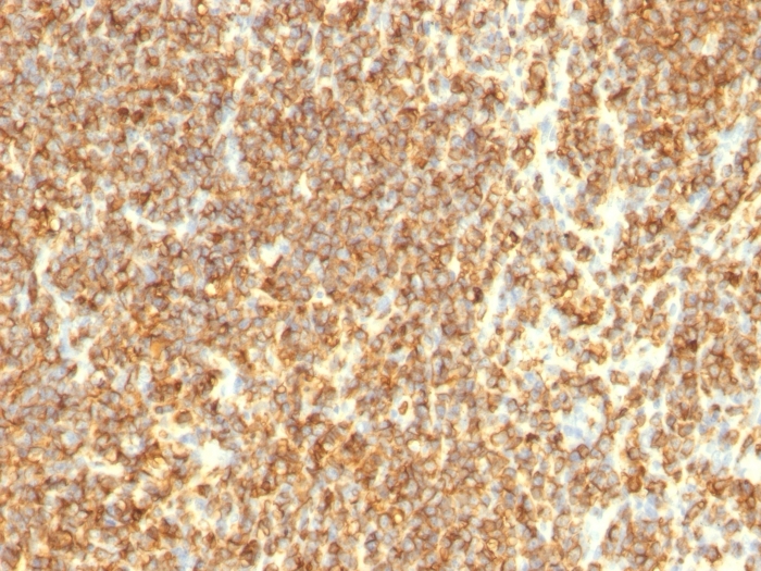
Formalin-fixed, paraffin-embedded human Lymphoma stained with CD20 Monoclonal Antibody (SPM494)
Formalin-fixed, paraffin-embedded human Lymphoma stained with CD20 Monoclonal Antibody (SPM494)
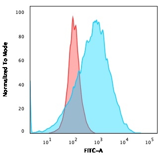
Flow Cytometric Analysis of Raji cells using CD20 Monoclonal Antibody (SPM494) followed by Goat anti-Mouse IgG-CF488 (Blue), Isotype Control (Red).
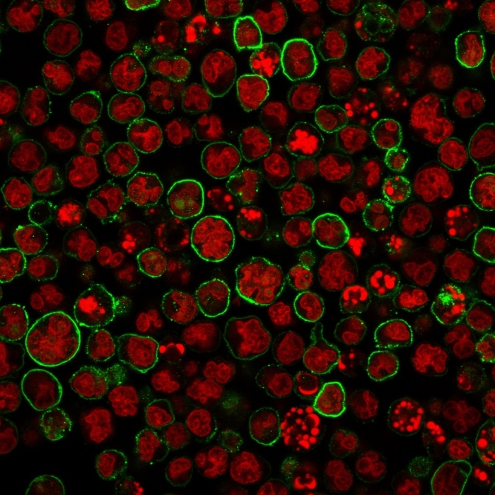
Immunofluorescence staining of MOLT-4 cells using CD20 Monoclonal Antibody (SPM494) followed by goat anti-Mouse IgG conjugated to CF488 (green). Nuclei are stained with Reddot.
* VAT and and shipping costs not included. Errors and price changes excepted