
YAP1 Antibody, KO Validated, Unconjugated, Rabbit, Polyclonal
Catalog Number:
PRS-13-379
- Images (9)
| Article Name: | YAP1 Antibody, KO Validated, Unconjugated, Rabbit, Polyclonal |
| Biozol Catalog Number: | PRS-13-379 |
| Supplier Catalog Number: | 13-379 |
| Alternative Catalog Number: | PRS-13-379-100 |
| Manufacturer: | ProSci |
| Host: | Rabbit |
| Category: | Antikörper |
| Application: | IF, IHC, IP, WB |
| Species Reactivity: | Human, Mouse, Rat |
| Immunogen: | Recombinant fusion protein containing a sequence corresponding to amino acids 155-504 of human YAP1 (NP_001123617.1). |
| Conjugation: | Unconjugated |
| Alternative Names: | YAP1, YAP, YAP2, YAP65, YKI |
| Application Dilute: | Optimal dilutions for each application to be determined by the researcher. |
| Application Notes: | WB: 1:500 - 1:2000IHC: 1:50 - 1:200IF: 1:50 - 1:200IP: 1:20 - 1:50 |
|
|
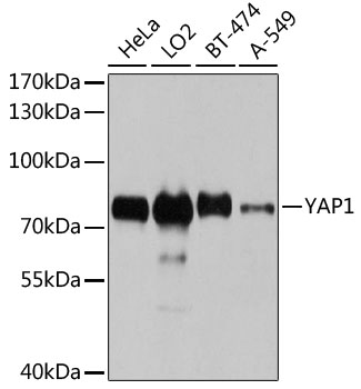
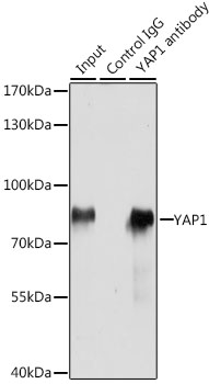
Western blot analysis of extracts of various cell lines, using YAP1 antibody (13-379) at 1:1000 dilution. Secondary antibody: HRP Goat Anti-Rabbit IgG (H+L) at 1:10000 dilution. Lysates/proteins: 25ug per lane. Blocking buffer: 3% nonfat dry milk in TBST. Detection: ECL Basic Kit. Exposure time: 10s. |
|
|
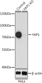
Western blot analysis of extracts from normal (control) and YAP1 knockout (KO) HeLa cells, using YAP1 antibody (13-379) at 1:1000 dilution. Secondary antibody: HRP Goat Anti-Rabbit IgG (H+L) at 1:10000 dilution. Lysates/proteins: 25ug per lane. Blocking buffer: 3% nonfat dry milk in TBST. Detection: ECL Enhanced Kit. Exposure time: 90s. |
|
|
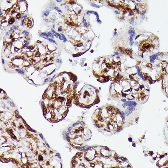
Immunohistochemistry of paraffin-embedded human placenta using YAP1 antibody (13-379) at dilution of 1:100 (40x lens). |
|
|
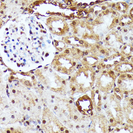
Immunohistochemistry of paraffin-embedded rat kidney using YAP1 antibody (13-379) at dilution of 1:100 (40x lens). |
|
|
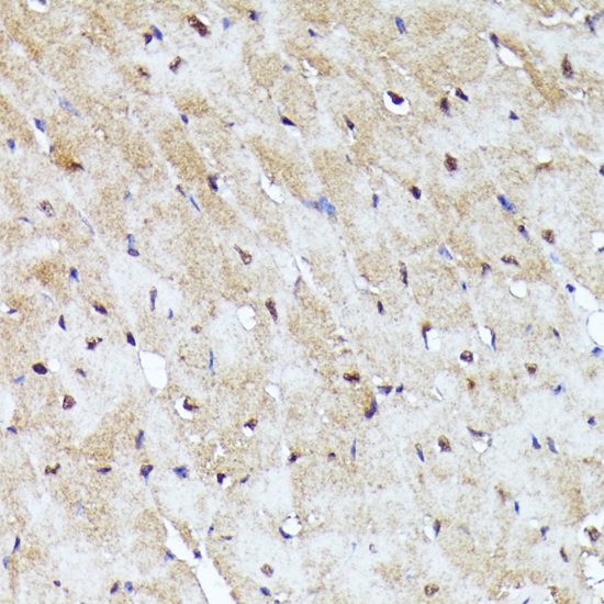
Immunohistochemistry of paraffin-embedded mouse heart using YAP1 antibody (13-379) at dilution of 1:100 (40x lens). |
|
|
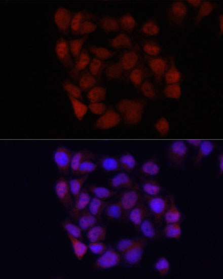
Immunofluorescence analysis of HeLa cells using YAP1 antibody (13-379) at dilution of 1:100. Blue: DAPI for nuclear staining. |
|
|
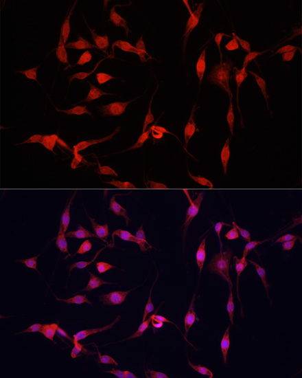
Immunofluorescence analysis of PC-12 cells using YAP1 antibody (13-370) at dilution of 1:100. Blue: DAPI for nuclear staining. |
|
|
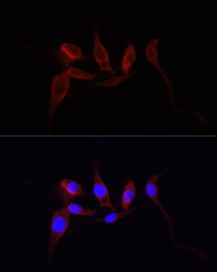
Immunofluorescence analysis of PC12 cells using YAP1 antibody (13-379) at dilution of 1:100. Blue: DAPI for nuclear staining. |
|
|
Immunoprecipitation analysis of 200ug extracts of HeLa cells, using 3 ug YAP1 antibody (13-379). Western blot was performed from the immunoprecipitate using YAP1 antibody (13-379) at a dilition of 1:1000. |
Product Guarantee and Expert Support
