
Acetyl-Histone H2B-K15 pAb, Unconjugated, Rabbit, Polyclonal
Catalog Number:
PRS-16-143
- Images (8)
| Article Name: | Acetyl-Histone H2B-K15 pAb, Unconjugated, Rabbit, Polyclonal |
| Biozol Catalog Number: | PRS-16-143 |
| Supplier Catalog Number: | 16-143 |
| Alternative Catalog Number: | PRS-16-143-100 |
| Manufacturer: | ProSci |
| Host: | Rabbit |
| Category: | Antikörper |
| Application: | IF, IHC, IP, WB |
| Species Reactivity: | Human, Mouse |
| Immunogen: | AA synthetic acetylated peptide corresponding to residues surrounding K15 of human Histone H2B |
| Conjugation: | Unconjugated |
| Clonality: | Polyclonal |
| Concentration: | batch dependent |
| Buffer: | PBS with 0.02% sodium azide, 50% glycerol, pH7.3. |
| Form: | Liquid |
| Application Dilute: | Optimal dilutions for each application to be determined by the researcher. |
| Application Notes: | WB: 1:500 - 1:2000IHC: 1:50 - 1:200IF: 1:50 - 1:100IP: 1:50 - 1:200 |
|
|
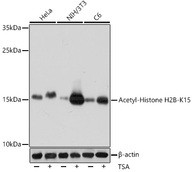
Western blot analysis of extracts of various cell lines, using Acetyl-Histone H2B-K15 antibody (16-143) at 1:1000 dilution.HeLa cells were treated by TSA (1 uM) at 37°C for 18 hours.NIH/3T3 cells were treated by TSA (1 uM) at 37°C for 18 hours.C6 cells were treated by TSA (1 uM) at 37°C for 18 hours. Secondary antibody: HRP Goat Anti-Rabbit IgG (H+L) at 1:10000 dilution. Lysates/proteins: 25ug per lane. Blocking buffer: 3% BSA. Detection: ECL Basic Kit. Exposure time: 10s. |
|
|
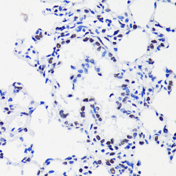
Immunohistochemistry of paraffin-embedded rat lung using H2B K15ac antibody (16-143) at dilution of 1:100 (40x lens). |
|
|
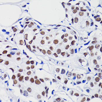
Immunohistochemistry of paraffin-embedded human breast cancer using H2B K15ac antibody (16-143) at dilution of 1:100 (40x lens). |
|
|
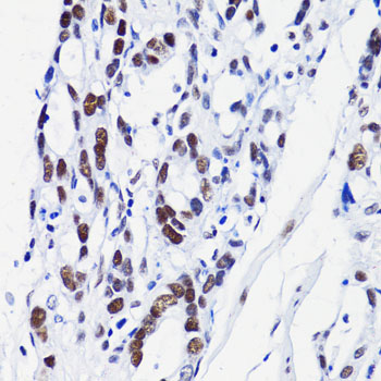
Immunohistochemistry of paraffin-embedded human gastric cancer using H2B K15ac antibody (16-143) at dilution of 1:100 (40x lens). |
|
|
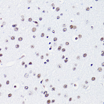
Immunohistochemistry of paraffin-embedded mouse brain using H2B K15ac antibody (16-143) at dilution of 1:100 (40x lens). |
|
|
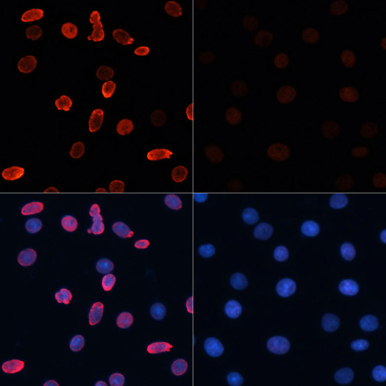
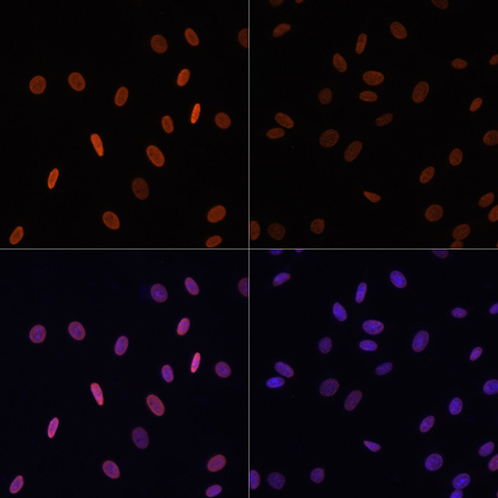
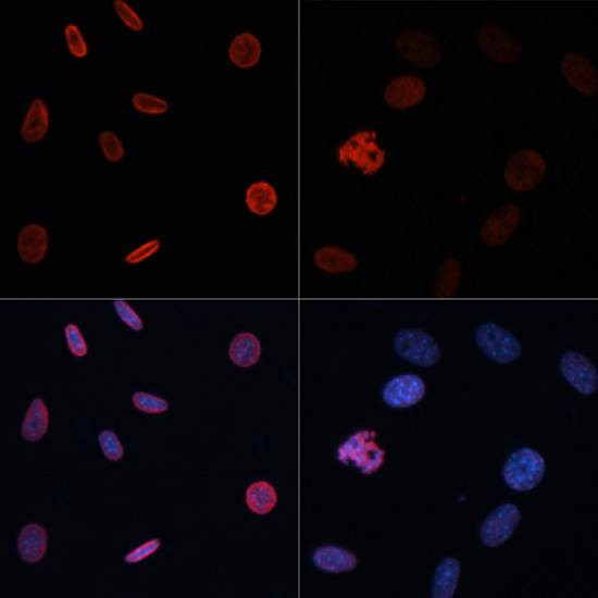
Immunofluorescence analysis of C6 cells using Acetyl-Histone H2B-K15 antibody (16-143) at dilution of 1:100.C6 cells were treated by TSA (1 uM) at 37°C for 18 hours. Blue: DAPI for nuclear staining. |
|
|
Immunofluorescence analysis of HeLa cells using Acetyl-Histone H2B-K15 antibody (16-143) at dilution of 1:100.HeLa cells were treated by TSA (1 uM) at 37°C for 18 hours. Blue: DAPI for nuclear staining. |
|
|
Immunofluorescence analysis of NIH/3T3 cells using Acetyl-Histone H2B-K15 antibody (16-143) at dilution of 1:100.NIH/3T3 cells were treated by TSA (1 uM) at 37°C for 18 hours. Blue: DAPI for nuclear staining. |
Product Guarantee and Expert Support
