
S100A4 Antibody, KO Validated, Unconjugated, Rabbit, Polyclonal
Catalog Number:
PRS-16-542
- Images (8)
| Article Name: | S100A4 Antibody, KO Validated, Unconjugated, Rabbit, Polyclonal |
| Biozol Catalog Number: | PRS-16-542 |
| Supplier Catalog Number: | 16-542 |
| Alternative Catalog Number: | PRS-16-542-100 |
| Manufacturer: | ProSci |
| Host: | Rabbit |
| Category: | Antikörper |
| Application: | IF, IHC, WB |
| Species Reactivity: | Human, Mouse, Rat |
| Immunogen: | Recombinant fusion protein containing a sequence corresponding to amino acids 1-101 of human S100A4 (NP_002952.1). |
| Conjugation: | Unconjugated |
| Alternative Names: | S100A4, S100 calcium binding protein A4, 18A2, 42A, CAPL, MTS1, P9KA, PEL98, S100 calcium binding protein A4 (calcium protein, calvasculin, metastasin, murine placental homolog), S100 calcium-binding protein A4, S100 calcium-binding protein A4 (calcium protein, malignant transformation suppression 1 |
| Application Dilute: | Optimal dilutions for each application to be determined by the researcher. |
| Application Notes: | WB: 1:500 - 1:2000IHC: 1:50 - 1:200IF: 1:50 - 1:200 |
|
|
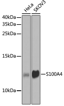
Western blot analysis of extracts of various cell lines, using S100A4 antibody (16-542) at 1:1000 dilution. Secondary antibody: HRP Goat Anti-Rabbit IgG (H+L) at 1:10000 dilution. Lysates/proteins: 25ug per lane. Blocking buffer: 3% nonfat dry milk in TBST. Detection: ECL Basic Kit. Exposure time: 90s. |
|
|
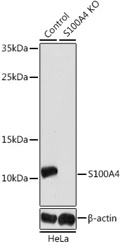
Western blot analysis of extracts from normal (control) and S100A4 knockout (KO) HeLa cells, using S100A4 antibody (16-542) at 1:1000 dilution. Secondary antibody: HRP Goat Anti-Rabbit IgG (H+L) at 1:10000 dilution. Lysates/proteins: 25ug per lane. Blocking buffer: 3% nonfat dry milk in TBST. Detection: ECL Enhanced Kit. Exposure time: 180s. |
|
|
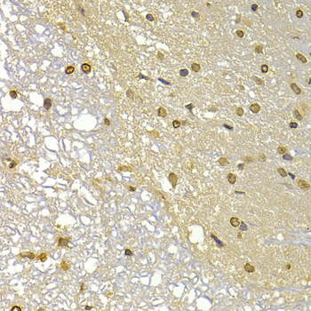
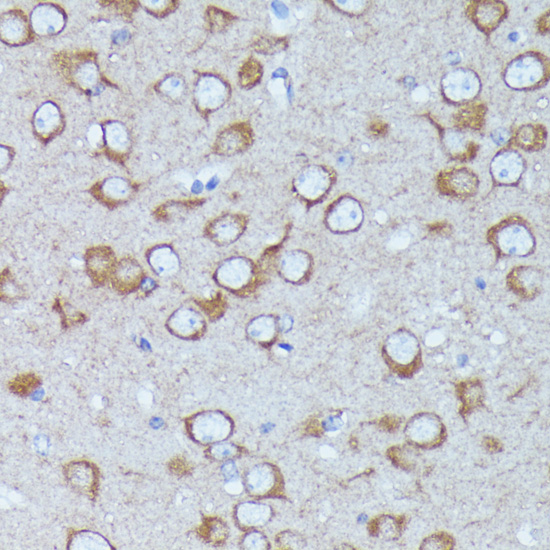
Immunohistochemistry of paraffin-embedded rat brain using S100A4 antibody (16-542) at dilution of 1:100 (40x lens). |
|
|
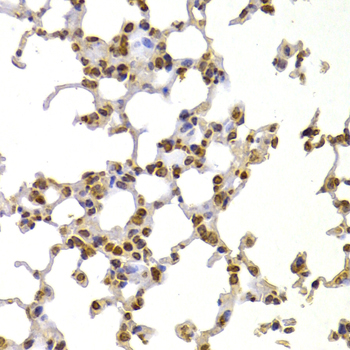
Immunohistochemistry of paraffin-embedded mouse lung using S100A4 antibody (16-542) at dilution of 1:100 (40x lens). |
|
|
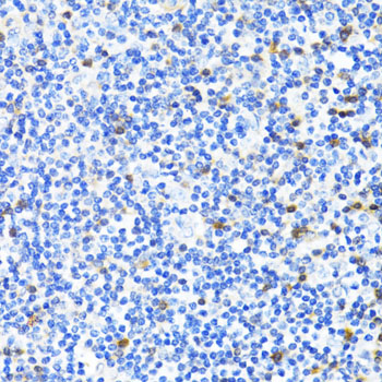
Immunohistochemistry of paraffin-embedded human tonsil using S100A4 antibody (16-542) at dilution of 1:100 (40x lens). |
|
|
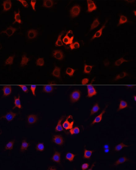
Immunofluorescence analysis of HeLa cells using S100A4 antibody (16-542) at dilution of 1:100. Blue: DAPI for nuclear staining. |
|
|
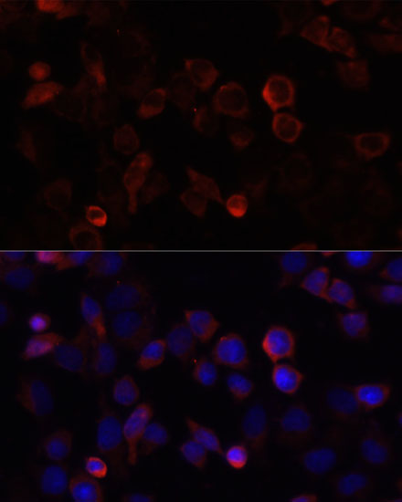
Immunofluorescence analysis of PC12 cells using S100A4 antibody (16-542) at dilution of 1:100. Blue: DAPI for nuclear staining. |
|
|
Immunofluorescence analysis of RAW264.7 cells using S100A4 antibody (16-542) at dilution of 1:100. Blue: DAPI for nuclear staining. |
Product Guarantee and Expert Support
