Optimal dilutions for each application to be determined by the researcher.
Application Notes:
WB: 1:500 - 1:2000, IHC: 1:50 - 1:200
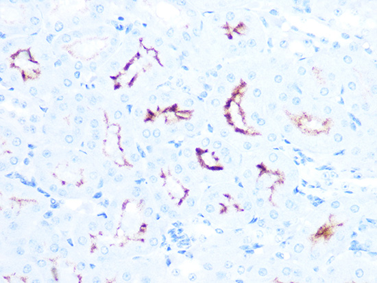
Figure 3 Immunohistochemistry Validation of ACE2 in Mouse Kidney Tissue Immunohistochemistry of paraffin-embedded mouse kidney tissue using ACE2 antibody, 24-033, at 1:100 dilution (40x lens).
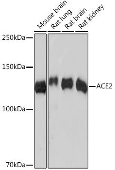
Figure 1 Western Blot Validation in Mouse and Rat Tissue LysatesLoading: 25 µg of lysates per lane.Antibodies: ACE2 antibody, 24-033, at 1:1000 dilution. Secondary: Goat anti-rabbit IgG HRP conjugate at 1:10000 dilution. Blocking buffer: 3% nonfat dry milk in TBST.
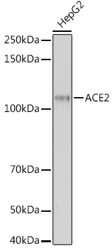
Figure 2 Western Blot Validation in HepG2 CellsLoading: 25 µg of lysates per lane. Antibodies: ACE2 antibody, 24-033, at 1:500 dilution. Secondary: Goat anti-rabbit IgG HRP conjugate at 1:10000 dilution. Blocking buffer: 3% nonfat dry milk in TBST.
* VAT and and shipping costs not included. Errors and price changes excepted