
IL-21 Antibody, Unconjugated, Rabbit, Polyclonal
Catalog Number:
PRS-2465
- Images (9)

| Article Name: | IL-21 Antibody, Unconjugated, Rabbit, Polyclonal |
| Biozol Catalog Number: | PRS-2465 |
| Supplier Catalog Number: | 2465 |
| Alternative Catalog Number: | PRS-2465-0.02,PRS-2465-0.1 |
| Manufacturer: | ProSci |
| Host: | Rabbit |
| Category: | Antikörper |
| Application: | ELISA, ICC, WB |
| Species Reactivity: | Human, Mouse, Rat |
| Immunogen: | Anti-IL-21 antibody (2465) was raised against a peptide corresponding to 15 amino acids near the center of human IL-21 precursor. The immunogen is located within the last 50 amino acids of IL-21. |
| Conjugation: | Unconjugated |
| Alternative Names: | IL-21 Antibody: Za11, IL-21, Interleukin-21, Za11 |
| Application Dilute: | Optimal dilutions for each application to be determined by the researcher. |
| Application Notes: | WB: 1 µg/mL, ICC: 2 µg/mL.Antibody validated: Western Blot in human, mouse and rat samples, Immunocytochemistry in human samples. All other applications and species not yet tested. |
|
|
Figure 3 Western Blot Validation in Human HL-60 Cell LysateLoading: 15 &956,g of lysates per lane.Antibodies: IL-21 2465 (1 &956,g/mL), 1h incubation at RT in 5% NFDM/TBST.Secondary: Goat anti-rabbit IgG HRP conjugate at 1:10000 dilution.(A) Absence of blocking peptide(B) Presence of blocking peptide. |
|
|
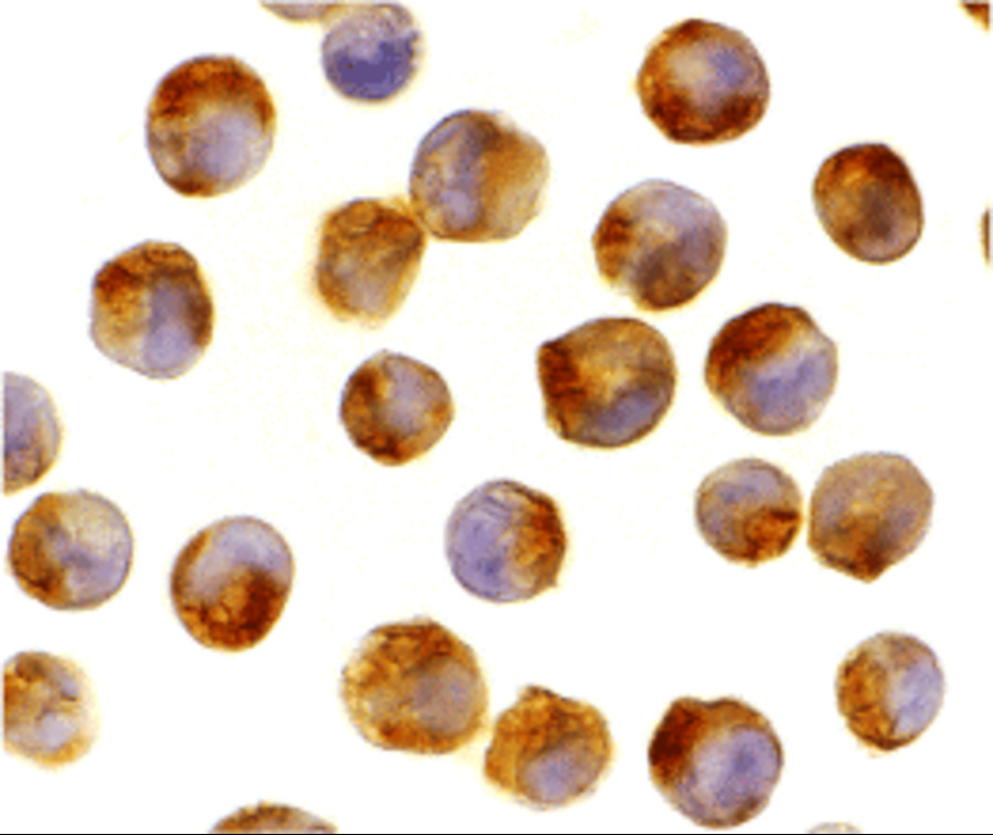
Figure 5 Immunocytochemistry Validation of IL-21 in K562 CellsImmunocytochemical analysis of K562 cells using anti-IL-21 antibody (2465) at 2 &956,g/mL. Cells was fixed with formaldehyde and blocked with 10% serum for 1 h at RT, antigen retrieval was by heat mediation with a citrate buffer (pH6). Samples were incubated with primary antibody overnight at 4&730, C. A goat anti-rabbit IgG H&L (HRP) at 1/250 was used as secondary. Counter stained with Hematoxylin. |
|
|
|
|
|
Figure 1 Western Blot Validation in Human, Mouse and Rat Cell LinesLoading: 15 &956,g of lysates per lane.Antibodies: IL-21 2465, (1 &956,g/mL), 1h incubation at RT in 5% NFDM/TBST.Secondary: Goat anti-rabbit IgG HRP conjugate at 1:10000 dilution. |
|
|
Figure 2 Independent Antibody Validation (IAV) via Protein Expression Profile in Cell LinesLoading: 15 &956,g of lysates per lane.Antibodies: IL-21 2465 (1 &956,g/mL), IL-21 2463 (5 &956,g/mL), beta-actin (5 &956,g/mL) and beta-actin (1 &956,g/mL), 1h incubation at RT in 5% NFDM/TBST.Secondary: Goat anti-rabbit IgG HRP conjugate at 1:10000 dilution. |
|
|
Figure 4 Western Blot Validation with Recombinant ProteinLoading: 30 ng of human IL-21 recombinant protein per lane.Antibodies: IL-21 2465 (0.5 &956,g/mL), 1h incubation at RT in 5% NFDM/TBST.Secondary: Goat anti-rabbit IgG HRP conjugate at 1:10000 dilution. |
|
|
Figure 6 Induced Expression Validation of IL-21 expresssion in patients with Crohns Disease (Monteleone et al, 2007) Enhanced IL-21 was observed in involved but not uninvolvedCD, and was not associated with any CD phenotype, such as fibrostenosing disease. IL-21 expression was detected by anti-IL-21 antibodies (2465). |
|
|
Figure 7 Regulation of IL-21 expresssion in duodenal biopsies of two patients with active Celiac disease (ACD) (Sarra et al., 2013) (b) shows IL-21 expression levels treated with dimethyl sulfoxide (DMSO) or wortmannin (WRT). (c) shows IL-21 expression levels with the inhibition of IL-15 antibody (aIL-15). IgG isotype was used as a control.Both WB show IL-21 expression decreased with the treatment with WRT or the inhibition of IL-15 antibody. |
|
|
Figure 8 Induced Expression Validation of IL-21 expresssion in patients with Helicobacter Pylori (Hp) (Caruso et al., 2007) (A) shows IL-21 expression levels from biopsies of patients with Hp infection and without Hp infection (n=3).(C) shows IL-21 expression levels in CD3+LPMC cells from Hp-positive and Hp-negative patients (n=3).Both WB show IL-21 expression increased in patients with Hp infection. |
Product Guarantee and Expert Support
