
PUMA Antibody, Unconjugated, Rabbit, Polyclonal
Catalog Number:
PRS-3041
- Images (9)
| Article Name: | PUMA Antibody, Unconjugated, Rabbit, Polyclonal |
| Biozol Catalog Number: | PRS-3041 |
| Supplier Catalog Number: | 3041 |
| Alternative Catalog Number: | PRS-3041-0.02,PRS-3041-0.1 |
| Manufacturer: | ProSci |
| Host: | Rabbit |
| Category: | Antikörper |
| Application: | ELISA, ICC, IF, WB |
| Species Reactivity: | Human, Mouse |
| Immunogen: | Anti-PUMA antibody (3041) was raised against a peptide corresponding to 14 amino acids near the carboxyl terminus human PUMA isoform 1. The immunogen is located within the last 50 amino acids of PUMA. |
| Conjugation: | Unconjugated |
| Alternative Names: | PUMA Antibody: JFY1, PUMA, JFY-1, Bcl-2-binding component 3 |
| Application Dilute: | Optimal dilutions for each application to be determined by the researcher. |
| Application Notes: | WB: 1-4 µg/mL, IF: 2 µg/mL, ICC: 1 µg/mL. Antibody validated: Western Blot in human and mouse samples, Immunocytochemistry and Immunofluorescence in human samples. All other applications and species not yet tested. |
|
|
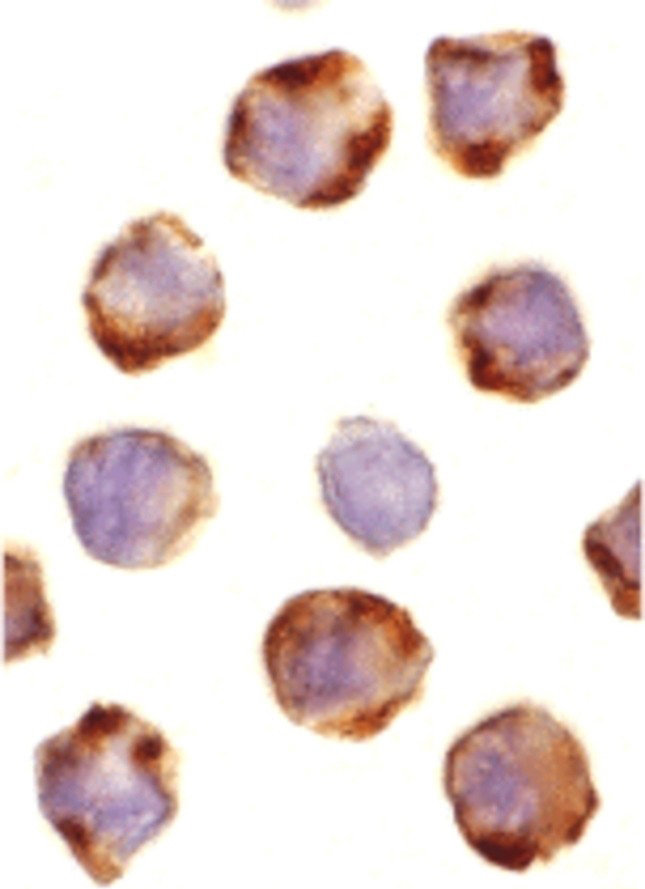
Figure 6 Immunocytochemistry Validation of PUMA in K562 Cells Immunocytochemical analysis of K562 cells using anti-PUMA antibody (3041) at 1 &956,g/ml. Cells was fixed with formaldehyde and blocked with 10% serum for 1 h at RT, antigen retrieval was by heat mediation with a citrate buffer (pH6). Samples were incubated with primary antibody overnight at 4&730,C. A goat anti-rabbit IgG H&L (HRP) at 1/250 was used as secondary. Counter stained with Hematoxylin. |
|
|
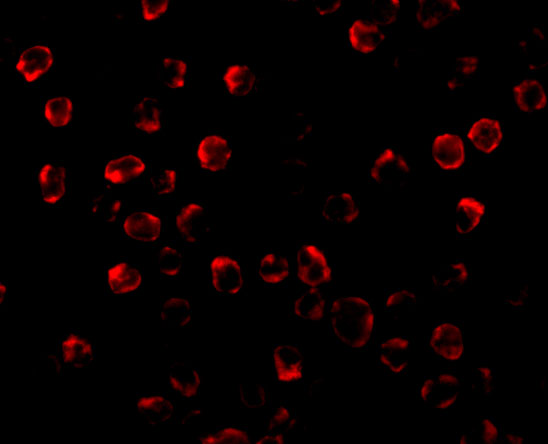
Figure 5 Immunofluorescence Validation of PUMA in K562 CellsImmunofluorescent analysis of 4% paraformaldehyde-fixed K562 cells labeling PUMA with 3041 at 2 &956,g/mL, followed by goat anti-rabbit IgG secondary antibody at 1/500 dilution (red). Image showing cytosol staining on K562 cells. |
|
|
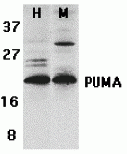
Figure 1 Western Blot Validation of PUMA in K562 and 3T3/NIH CellsLoading: 15 &956,g of lysates per lane.Antibodies: 3041 (2 &956,g/mL), 1 h incubation at RT in 5% NFDM/TBST.Secondary: Goat anti-rabbit IgG HRP conjugate at 1:10000 dilution. |
|
|
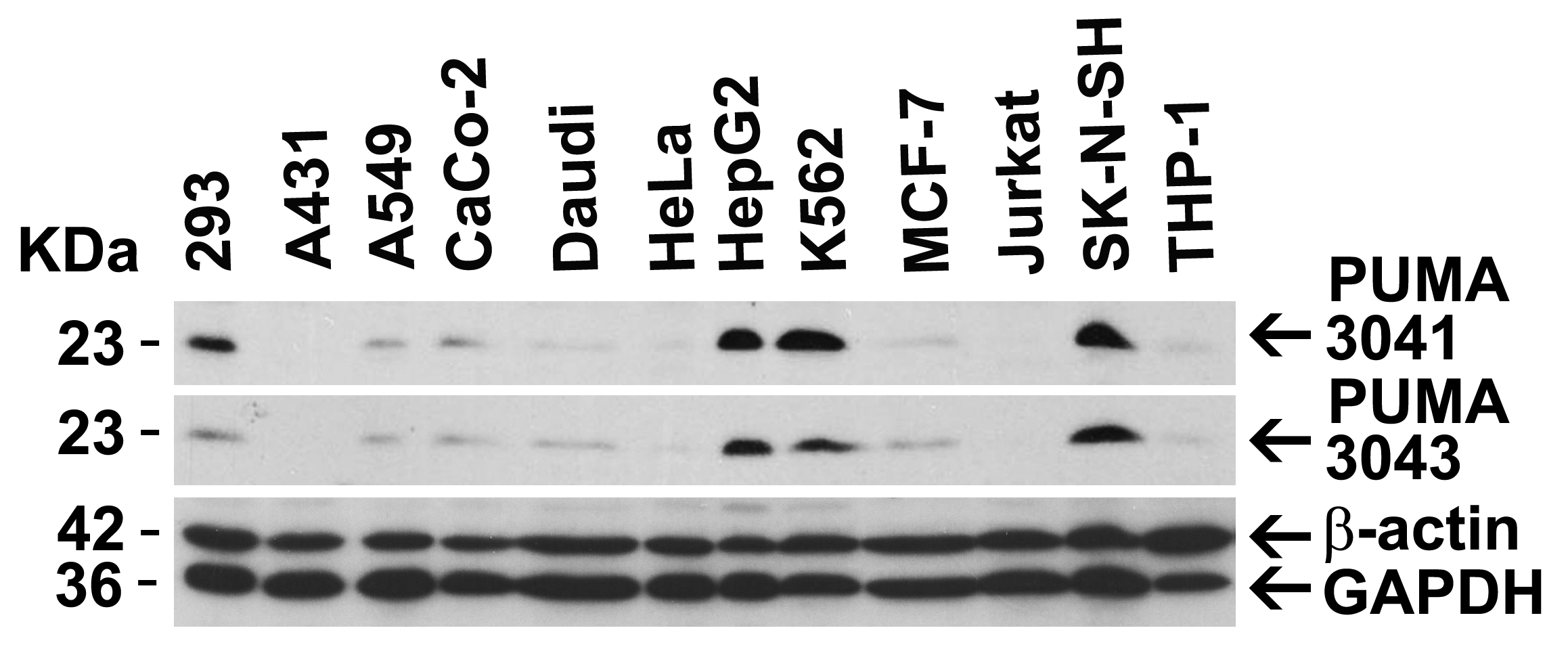
Figure 2 Independent Antibody Validation (IAV) via Protein Expression Profile in Cell LinesLoading: 20 &956,g of lysates per lane.Antibodies: 3041 (3 &956,g/mL), 3043 (2 &956,g/mL), beta-actin (1 &956,g/mL) and GAPDH (0.02 &956,g/mL), 1 h incubation at RT in 5% NFDM/TBST.Secondary: Goat anti-rabbit IgG HRP conjugate at 1:10000 dilution. |
|
|
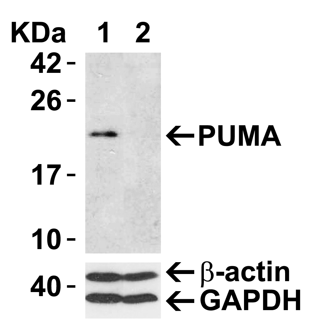
Figure 3 Validation with PUMA siRNA Knockdown in 293 Cells 293 cells were transfected with control siRNAs (lane 1) or PUMA siRNAs (lane 2) Loading: 15 &956,g of 293 whole cell lysates per lane.Antibodies: 3041 (2 &956,g/mL), beta-actin (1 &956,g/mL) and GAPDH (0.02 &956,g/mL), 1 h incubation at RT in 5% NFDM/TBST.Secondary: Goat anti-rabbit IgG HRP conjugate at 1:10000 dilution. |
|
|
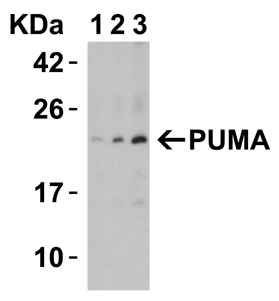
Figure 4 Sensitivity Test for PUMA in 2983 Cells Loading: Lysates/proteins at 15 &956,g per lane.Antibodies: 3041 (lane 1-3: 1, 2 and 4 &956,g/mL). 1 h incubation at RT in 5% NFDM/TBST.Secondary: Goat anti-rabbit IgG HRP conjugate at 1:10000 dilution. |
|
|
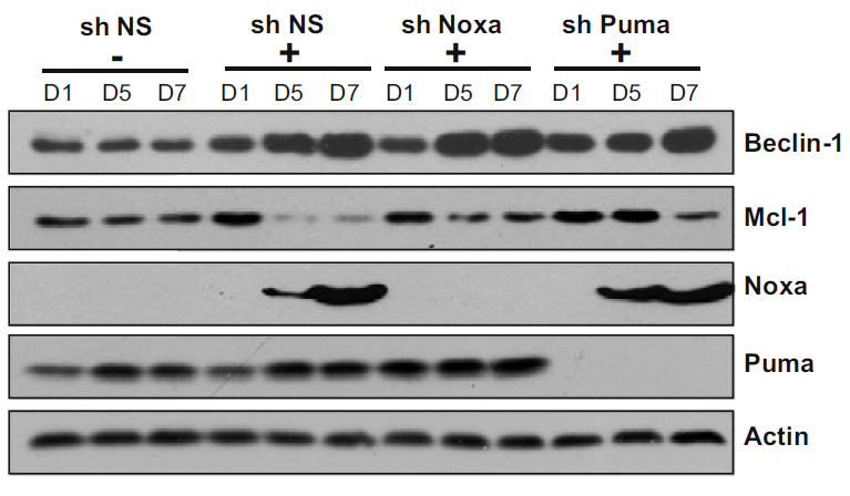
Figure 7 KD Validation of PUMA in HOSE-RasV12 Cells (Elgendy et al., 2011) HOSE-RasV12 cells were transfected with control shRNA plasmid or shRNA plasmids (KD) targeted against Noxa or Puma, as indicated. PUMA expression was not observed in PUMA KD cells detected by anti-PUMA antibodies (3041). |
|
|
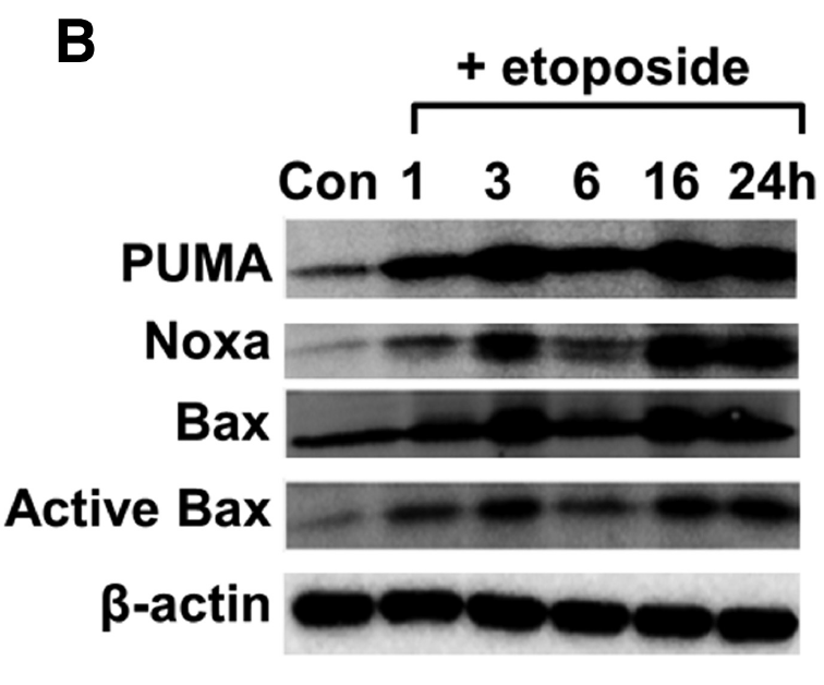
Figure 8 Induction Validation of PUMA in Primary Cortical Neurons (Sabirzhanov et al., 2014) PUMA protein levels were increased in etoposide-treated primary cortical neurons detected by anti-PUMA antibodies (3041). |
|
|
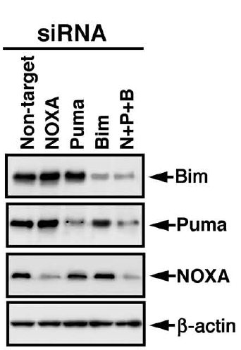
Figure 9 KD Validation of PUMA PUMA in Tet Cells (Han et al., 2010) Immunoblot analyses of Tet-induced p53 cells treated with NOXA, Puma, Bim or non-targeting siRNAs that were utilized in this experiment. PUMA protein levels were markedly reduced in PUMA KD cells detected by anti-PUMA antibodies (3041). |
Product Guarantee and Expert Support
