
PHAP I Antibody, Unconjugated, Rabbit, Polyclonal
Catalog Number:
PRS-3145
- Images (9)
| Article Name: | PHAP I Antibody, Unconjugated, Rabbit, Polyclonal |
| Biozol Catalog Number: | PRS-3145 |
| Supplier Catalog Number: | 3145 |
| Alternative Catalog Number: | PRS-3145-0.02,PRS-3145-0.1 |
| Manufacturer: | ProSci |
| Host: | Rabbit |
| Category: | Antikörper |
| Application: | ELISA, ICC, IF, WB |
| Species Reactivity: | Human, Mouse, Rat |
| Immunogen: | Anti-PHAP I antibody (3145) was raised against a peptide corresponding to 15 amino acids near the carboxy terminus of human PHAP I. The immunogen is located within the last 50 amino acids of PHAP I. |
| Conjugation: | Unconjugated |
| Alternative Names: | PHAP I Antibody: LANP, MAPM, PP32, HPPCn, PHAP1, PHAPI, I1PP2A, C15orf1, LANP, Acidic leucine-rich nuclear phosphoprotein 32 family member A, Acidic nuclear phosphoprotein pp32 |
| Application Dilute: | Optimal dilutions for each application to be determined by the researcher. |
| Application Notes: | WB: 2-4 µg/mL, ICC: 2 µg/mL, IF: 10 µg/mL.Antibody validated: Western Blot in human, mouse and rat samples, Immunocytochemistry in human samples, Immunofluorescence in human samples. All other applications and species not yet tested. |
|
|
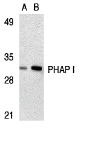
Figure 1 Western Blot Validation in Human Raji Cell LysateLoading: 15 &956,g of lysates per lane.Antibodies: PHAP I 3145 (A: 2 &956,g/mL, B: 4 &956,g/mL), 1h incubation at RT in 5% NFDM/TBST.Secondary: Goat anti-rabbit IgG HRP conjugate at 1:10000 dilution. |
|
|
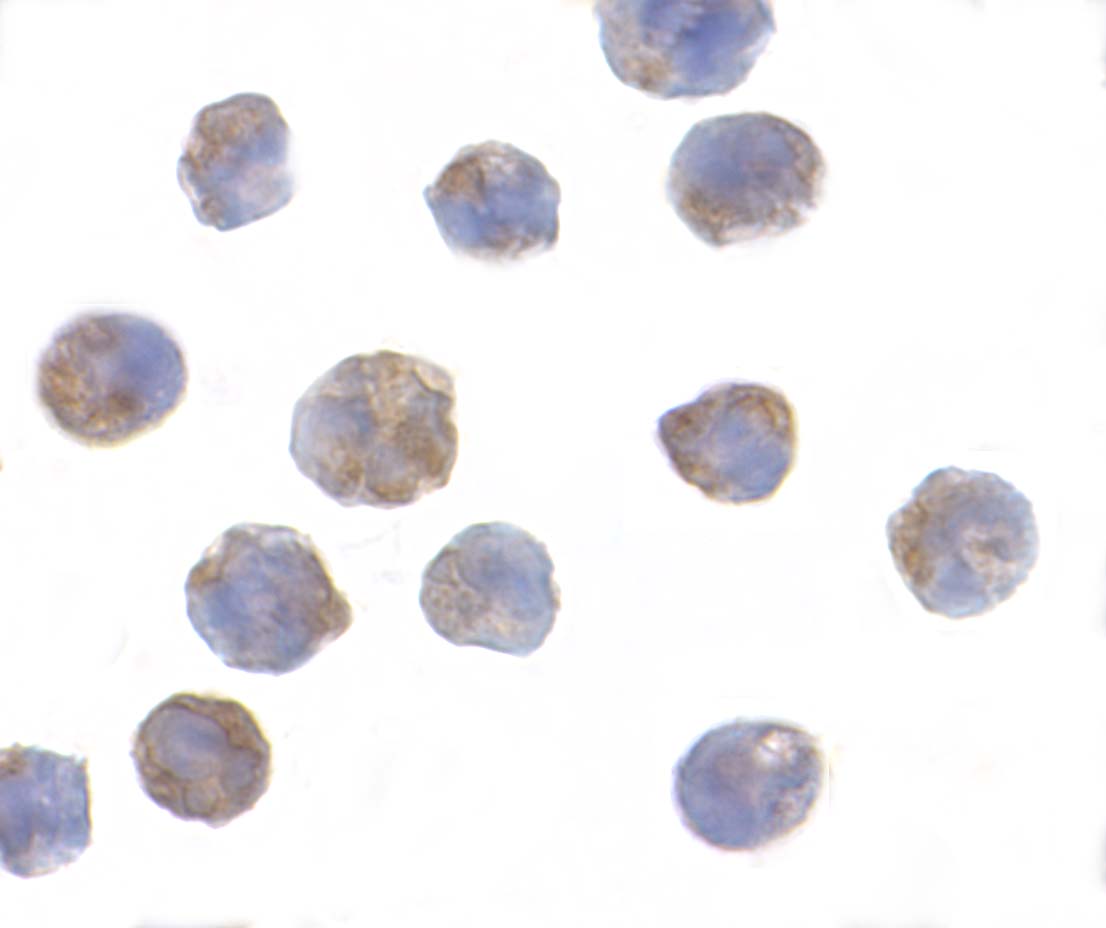
Figure 5 Immunocytochemistry Validation of PHAP I in Raji CellsImmunocytochemical analysis of Raji cells using anti-PHAP I antibody (3145) at 2 &956,g/ml. Cells was fixed with formaldehyde and blocked with 10% serum for 1 h at RT, antigen retrieval was by heat mediation with a citrate buffer (pH6). Samples were incubated with primary antibody overnight at 4&730,C. A goat anti-rabbit IgG H&L (HRP) at 1/250 was used as secondary. Counter stained with Hematoxylin. |
|
|
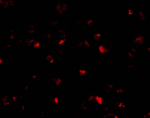
Figure 4 Immunofluorescence Validation of PHAP I in Raji CellsImmunofluorescent analysis of 4% paraformaldehyde-fixed Raji Cells labeling PHAP I with 3145 at 10 &956,g/mL, followed by goat anti-rabbit IgG secondary antibody at 1/500 dilution (red). |
|
|
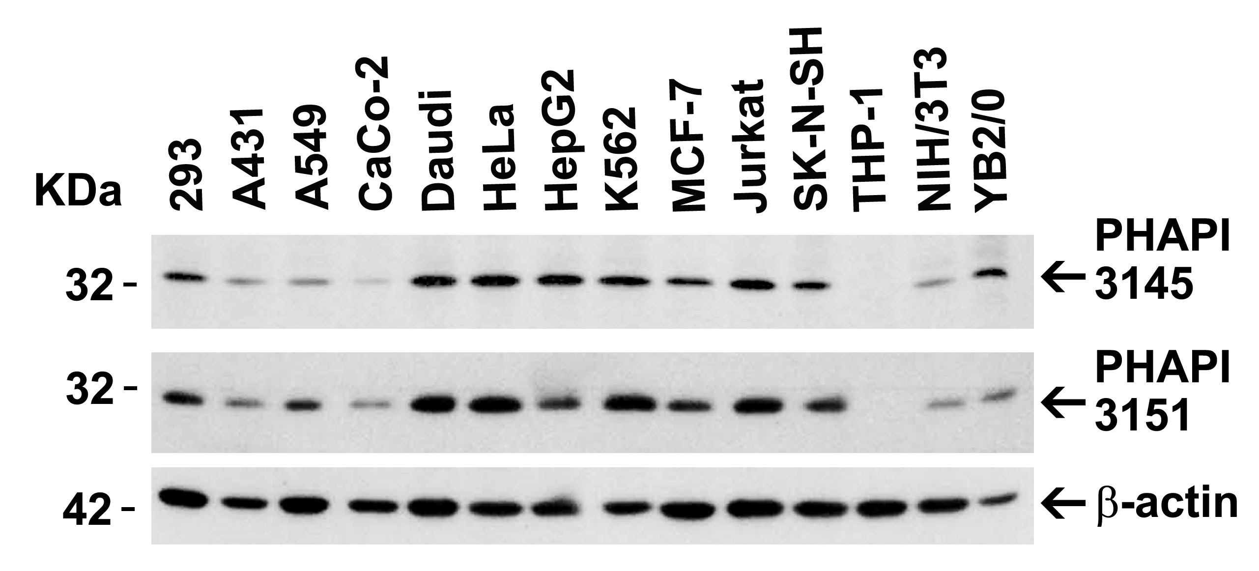
Figure 2 Independent Antibody Validation (IAV) via Protein Expression Profile in Cell LinesLoading: 15 &956,g of lysates per lane.Antibodies: PHAP I 3145 (2 &956,g/mL), PHAP I 3151 (1 &956,g/mL), and beta-actin (1 &956,g/mL), 1h incubation at RT in 5% NFDM/TBST.Secondary: Goat anti-rabbit IgG HRP conjugate at 1:10000 dilution. |
|
|
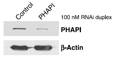
Figure 6 KD Validation of PHAPI in Human Breast Cancer Cells (Schafer et al., 2006) Human Breast Cancer Cells (T47D cells) were transfected with control or PHAPI siRNA duplex. PHAPI was detected via Western Blot analysis by using the anti-PHAPI antibody. PHAPI expression was reduced after PHAPI siRNA knockdown. |
|
|
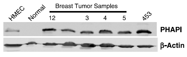
Figure 7 Increased Expression Validation of PHAPI in Patient Samples of BreastTumor Tissue (Schafer et al., 2006) PHAPI was overexpressed in all breast tumor samples of patients and human breast cancer cells (MDA-MB-453), but not in the normal breast tissue or human primary mammary epithelial cells (HMEC). |
|
|
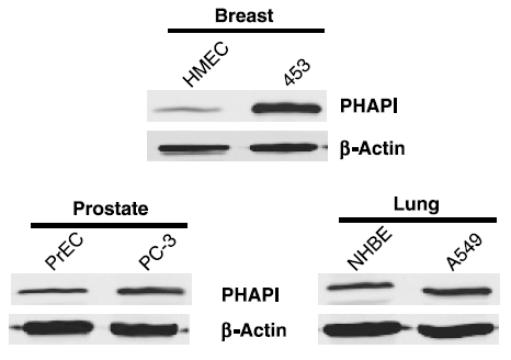
Figure 8 Overexpression of PHAPI in Breast Cancer Cells (Schafer et al., 2006) Western blot analysis with anti-PHAPI antibodies was performed for PHAPI in human cell lines from breast, prostate and lung. PHAPI was overexpressed in breast cancer cells when compared with normal cells (HMEC) whereas there were no significant differences in PHAPI expression in normal and cancer cells of either prostate or lung origin. |
|
|
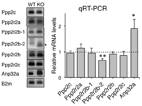
Figure 9 Induced Expression Validation of PHAPI/Anp32a in Atxn1 KO Mice (Sa´nchez et al., 2013) Western blot analysis of PHAPI/Anp32a from the cerebellum of WT and Atxn1 KO mice. PHAPI expression was significantly increased (2 folds) in Atxn1 KO mice as compared to WT mice. The same effect was observed in PHAPI mRNA levels. |
|
|
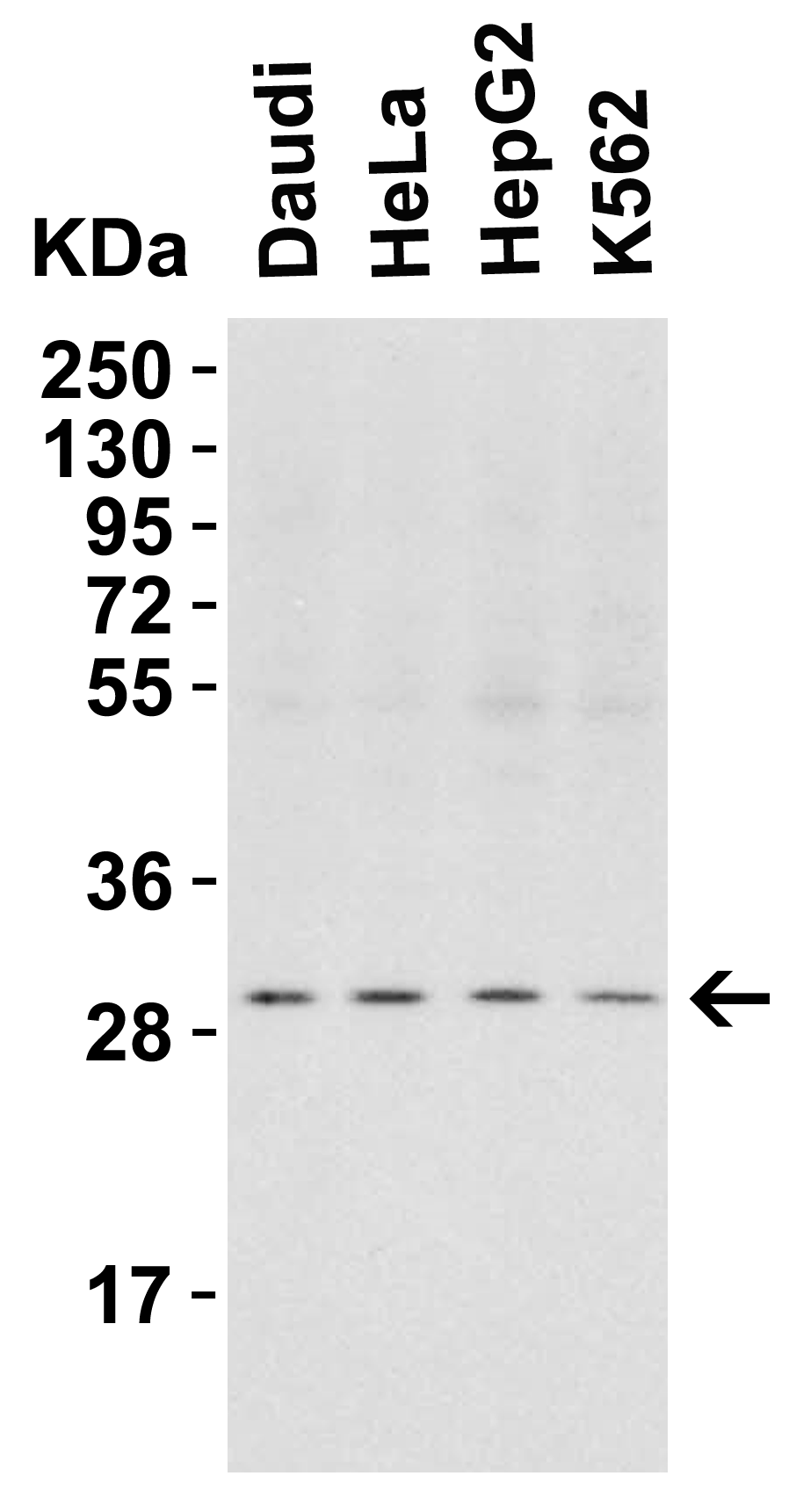
Figure 3 Western Blot Validation in Human Cell LinesLoading: 15 &956,g of lysates per lane.Antibodies: PHAP I 3145 (2 &956,g/mL), 1h incubation at RT in 5% NFDM/TBST.Secondary: Goat anti-rabbit IgG HRP conjugate at 1:10000 dilution. |
Product Guarantee and Expert Support
