
TIP47 Antibody, Unconjugated, Rabbit, Polyclonal
Catalog Number:
PRS-3883
- Images (9)
| Article Name: | TIP47 Antibody, Unconjugated, Rabbit, Polyclonal |
| Biozol Catalog Number: | PRS-3883 |
| Supplier Catalog Number: | 3883 |
| Alternative Catalog Number: | PRS-3883-0.02,PRS-3883-0.1 |
| Manufacturer: | ProSci |
| Host: | Rabbit |
| Category: | Antikörper |
| Application: | ELISA, IHC-P, WB |
| Species Reactivity: | Human, Mouse |
| Immunogen: | Anti-TIP47 antibody (3883) was raised against a peptide corresponding to 14 amino acids near the amino terminus of human TIP47. The immunogen is located within amino acids 30 - 80 of TIP47. |
| Conjugation: | Unconjugated |
| Alternative Names: | TIP47 Antibody: PP17, TIP47, M6PRBP1, Perilipin-3, 47 kDa mannose 6-phosphate receptor-binding protein, 47 kDa MPR-binding protein |
| Application Dilute: | Optimal dilutions for each application to be determined by the researcher. |
| Application Notes: | WB: 1-4 µg/mL, IHC-P: 2 µg/mL.Antibody validated: Western Blot in human and mouse samples, Immunohistochemistry in rat samples. All other applications and species not yet tested. |
|
|
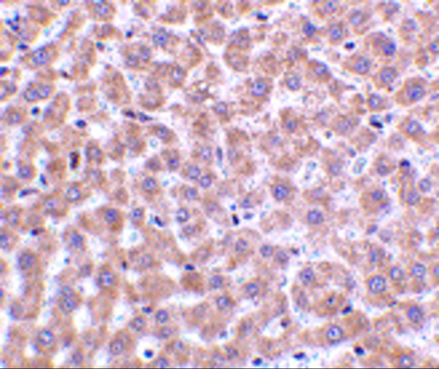
Figure 5 Immunohistochemistry Validation of TIP47 in Rat Liver Tissue Immunohistochemical analysis of paraffin-embedded rat liver tissue using anti-TIP47 antibody (3883) at 2 &956,g/ml. Tissue was fixed with formaldehyde and blocked with 10% serum for 1 h at RT, antigen retrieval was by heat mediation with a citrate buffer (pH6). Samples were incubated with primary antibody overnight at 4&730,C. A goat anti-rabbit IgG H&L (HRP) at 1/250 was used as secondary. Counter stained with Hematoxylin. |
|
|
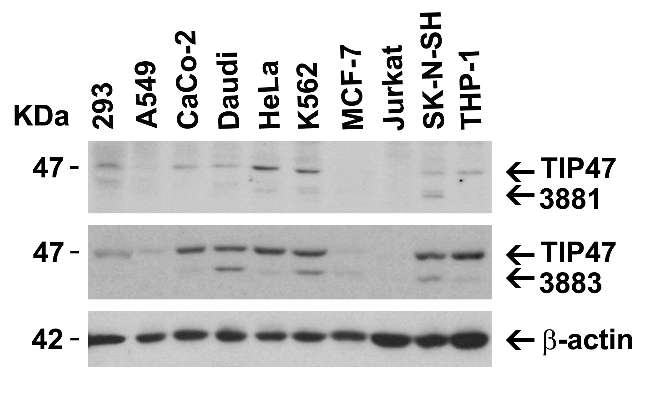
Figure 1 Western Blot Validation in Human Cell LinesLoading: 15 &956,g of lysates per lane.Antibodies: TIP47 3883, (1 µg/mL), 1h incubation at RT in 5% NFDM/TBST.Secondary: Goat anti-rabbit IgG HRP conjugate at 1:10000 dilution |
|
|
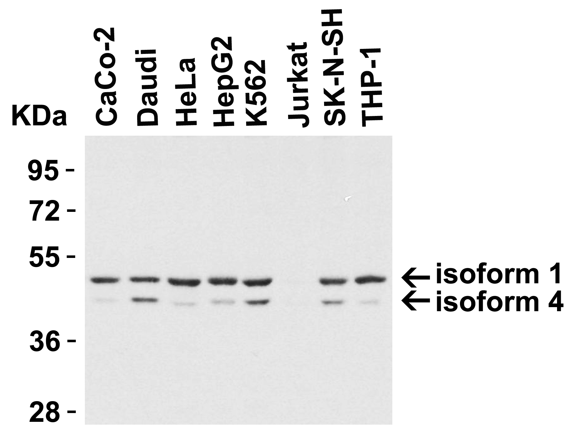
Figure 2 Independent Antibody Validation (IAV) via Protein Expression Profile in Human Cell LinesLoading: 15 µg of lysates per lane.Antibodies: TIP47 3881 (4 &956,g/mL), TIP47 3883 (1 &956,g/mL), and beta-actin (1 &956,g/mL), 1h incubation at RT in 5% NFDM/TBST.Secondary: Goat anti-rabbit IgG HRP conjugate at 1:10000 dilution. |
|
|
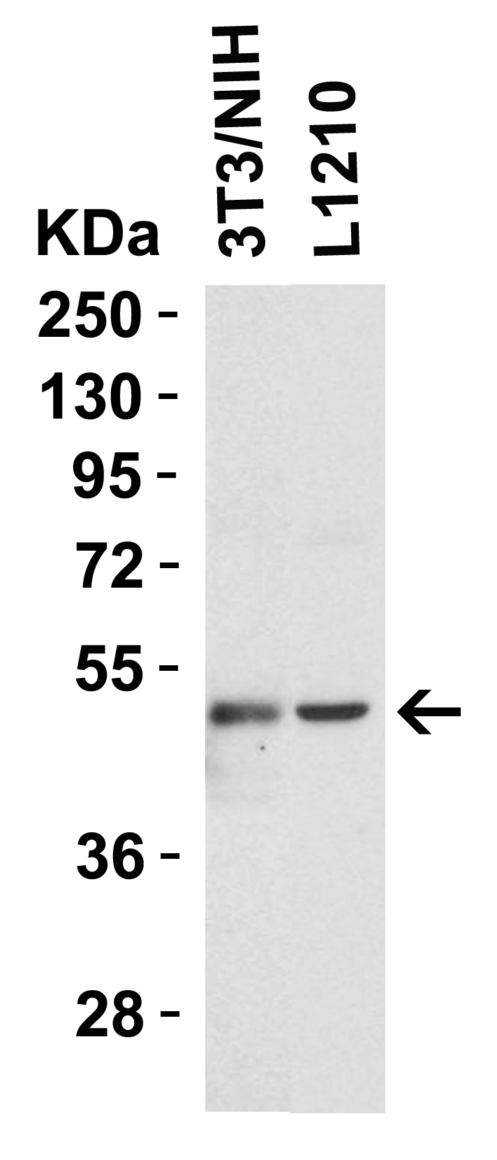
Figure 3 Western Blot Validation in Mouse Cell LinesLoading: 15 &956,g of lysates per lane.Antibodies: TIP47 3883 (1 &956,g/mL), 1h incubation at RT in 5% NFDM/TBST.Secondary: Goat anti-rabbit IgG HRP |
|
|
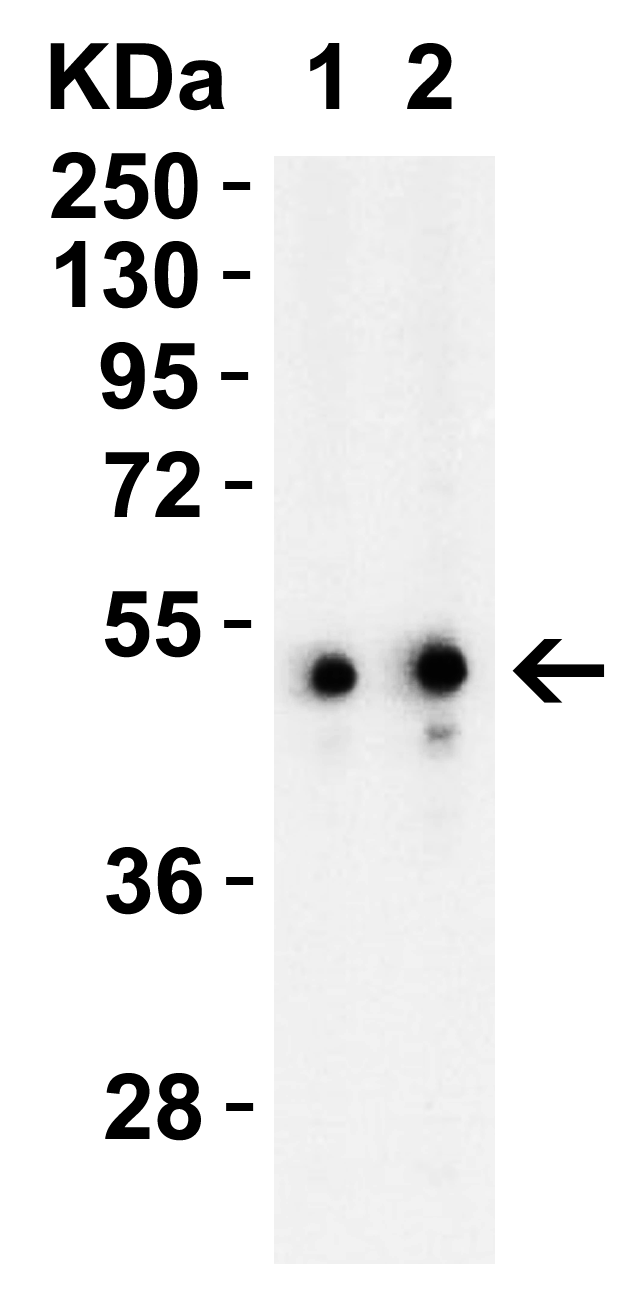
Figure 4 Western Blot Validation in Mouse EL4 CellsLoading: 15 &956,g of lysates per lane.Antibodies: TIP47 3883, 1h incubation at RT in 5% NFDM/TBST.Secondary: Goat anti-rabbit IgG HRP conjugate at 1:10000 dilution.Lane 1: 1 &956,g/mL Lane 2: 2 &956,g/mL |
|
|
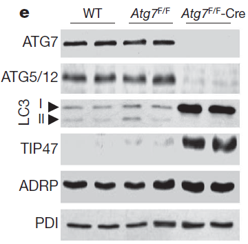
Figure 6 Induced Expression Validation of TIP47 in Atg7 Liver KO Mice (Singh et al., 2009) TIP47 expression detected by anti-TIP47 antibodies was markedly increased in the liver of Atg7 liver KO mice. |
|
|
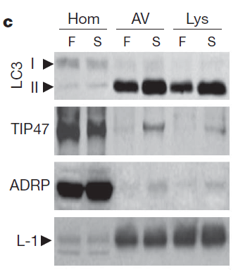
Figure 7 Induced Expression Validation of TIP47 in Mouse Liver (Singh et al., 2009) TIP47 expression detected by anti-TIP47 antibodies was increased in the autophagic vacuoles (AVs) and lysosomes (Lys) after starvation. |
|
|
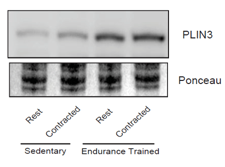
Figure 8 Induced Expression Validation of TIP47 in Rat Skeletal Muscle (Ramos et al., 2015) Mitochondrial red gastrocnemius muscle PLIN3 (TIP47) content was increased in endurance-trained rats as compared to sedentary rats. PLIN3 protein was detected by anti-TIP47 antibodies. |
|
|
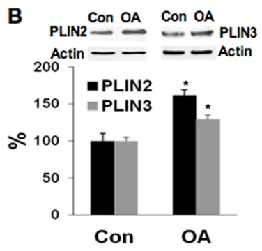
Figure 9 Induced Expression Validation of TIP47 in HT29 Cells (Qi et al., 2013) TIP47 expression detected by anti-TIP47 antibodies was increased in the colonic cells after oleic acid treatment. |
Product Guarantee and Expert Support
