
SCF Antibody, Unconjugated, Rabbit, Polyclonal
Catalog Number:
PRS-5165
- Images (7)


| Article Name: | SCF Antibody, Unconjugated, Rabbit, Polyclonal |
| Biozol Catalog Number: | PRS-5165 |
| Supplier Catalog Number: | 5165 |
| Alternative Catalog Number: | PRS-5165-0.02,PRS-5165-0.1 |
| Manufacturer: | ProSci |
| Host: | Rabbit |
| Category: | Antikörper |
| Application: | ELISA, IF, IHC-P, WB |
| Species Reactivity: | Human, Mouse, Rat |
| Immunogen: | Anti-SCF antibody (5165) was raised against a peptide corresponding to 18 amino acids near the center of human SCF. The immunogen is located within amino acids 100-150 of SCF. |
| Conjugation: | Unconjugated |
| Alternative Names: | SCF Antibody: SF, MGF, SCF, FPH2, KL-1, Kitl, SHEP7, Kit ligand, Mast cell growth factor |
| Application Dilute: | Optimal dilutions for each application to be determined by the researcher. |
| Application Notes: | WB: 1-2 µg/mL, IHC: 2.5 µg/mL, IF: 20 µg/mL.Antibody validated: Western Blot in human, mouse and rat samples, Immunohistochemistry in mouse samples, Immunofluorescence in human samples. All other applications and species not yet tested. |
|
|
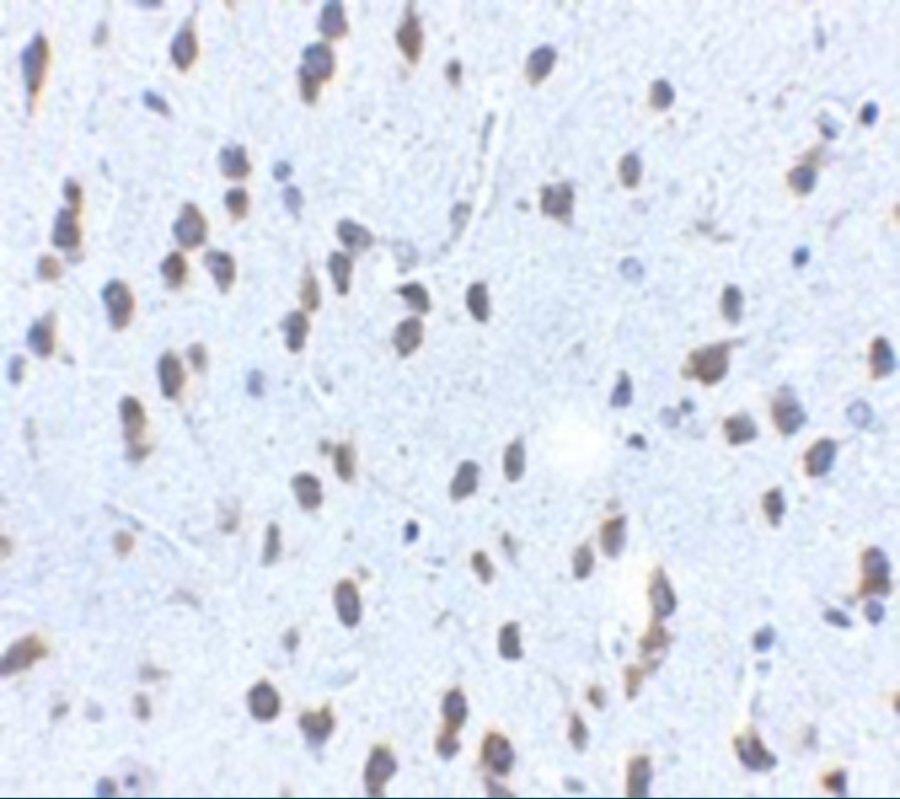
Immunohistochemistry Validation of SCF in Mouse Brain Tissue Immunohistochemical analysis of paraffin-embedded mouse brain tissue using anti-SCF antibody (5165) at 2.5 &956,g/ml. Tissue was fixed with formaldehyde and blocked with 10% serum for 1 h at RT, antigen retrieval was by heat mediation with a citrate buffer (pH6). Samples were incubated with primary antibody overnight at 4 &730,C. A goat anti-rabbit IgG H&L (HRP) at 1/250 was used as secondary. Counter stained with Hematoxylin. |
|
|
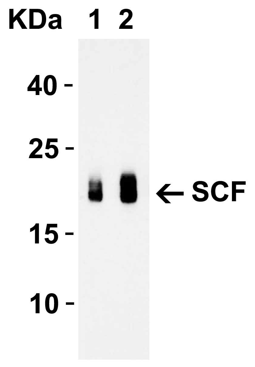
Western Blot Validation with Recombinant ProteinLoading: 30 ng of human SCF recombinant protein per lane.Antibodies: SCF 5165 (Lane 1: 1 &956,g/mL and Lane 2: 2 &956,g/mL), 1h incubation at RT in 5% NFDM/TBST.Secondary: Goat anti-rabbit IgG HRP conjugate at 1:10000 dilution.Observed at around 20kD. |
|
|
Immunofluorescence Validation of SCF in Human Brain TissueImmunofluorescent analysis of 4% paraformaldehyde-fixed human brain tissue labeling SCF with 5165 at 20 &956,g/mL, followed by goat anti-rabbit IgG secondary antibody at 1/500 dilution (red). |
|
|
Western Blot Validation in Cell Lines and Tissues of Human, Mouse and RatLoading: 15 &956,g of lysates per lane.Antibodies: SCF 5165 (1 &956,g/mL), 1h incubation at RT in 5% NFDM/TBST.Secondary: Goat anti-rabbit IgG HRP conjugate at 1:10000 dilution. |
|
|
Regulation Validation of SCF in Streptozotocin (STZ)-induced Diabetic Mice (Zhang et al., 2016) WB analysis showed protein expression level of SCF detected by anti-SCF antibody (5165) in gastric smooth muscle was significantly decreased in STZ-induced diabetic mice as compared to the control. |
|
|
Regulated Expression Validation of SCF in Gastric Smooth Muscle Cells (GSMCs) of Mice (Zhang et al., 2016) WB analysis of protein expression level of SCF detected by anti-SCF antibody (5165). GSMCs were treated with PI3K inhibitor (LY294002), AT1R inhibitor (ZD7155) and AT2R inhibitor (PD123319) before Ang II treatment. Ang II (10-8mol/L) significantly increased SCF protein expression, which was reduced by treatment of LY294002 and ZD7155. |
|
|
Regulated Expression Validation of SCF in GSMCs of Normal Mice (Wu et al., 2013) WB analysis of protein expression level of SCF detected by anti-SCF antibody (5165). GSMCs were treated with C-type natriuretic peptide (CNP) at different doses for 48hr. CNP(10-7mol/L and 10-6mol/L) significantly decreased SCF protein expression level as compared to the control group. |
Product Guarantee and Expert Support
