
LMX1A Antibody, Unconjugated, Rabbit, Polyclonal
Catalog Number:
PRS-7087
- Images (7)
| Article Name: | LMX1A Antibody, Unconjugated, Rabbit, Polyclonal |
| Biozol Catalog Number: | PRS-7087 |
| Supplier Catalog Number: | 7087 |
| Alternative Catalog Number: | PRS-7087-0.02,PRS-7087-0.1 |
| Manufacturer: | ProSci |
| Host: | Rabbit |
| Category: | Antikörper |
| Application: | ELISA, ICC, IHC-P, WB |
| Species Reactivity: | Human, Mouse, Rat |
| Immunogen: | Rabbit polyclonal LMX1A antibody was raised against a 16 amino acid peptide near the carboxy terminus of human LMX1A.The immunogen is located within amino acids 300 - 350 of LMX1A. |
| Conjugation: | Unconjugated |
| Alternative Names: | LMX1A Antibody: LMX1, LMX1.1, LIM homeobox transcription factor 1-alpha |
| Application Dilute: | Optimal dilutions for each application to be determined by the researcher. |
| Application Notes: | WB: 1-2 µg/mL, ICC/IF: 10-20 µg/mL, IHC-P: 1-5 µg/mL. Antibody validated: Western Blot in human, mouse and rat samples, Immunocytochemistry in human samples, Immunohistochemistry in human, mouse, and rat samples. All other applications and species not yet tested. |
|
|
Figure 2 Western Blot Validation in Human Skeletal Muscle Loading: 10 &956,g of lysates per lane.A&956,g ntibodies: LMX1A, 7087, 1 &956,g/mL, 1h incubation at RT in 5% NFDM/TBST.Secondary: Goat anti-rabbit IgG HRP conjugate at 1:10000 dilution. |
|
|
Figure 3 Western Blot Validation in Mouse Skeletal Muscle Loading: 10 &956,g of lysates per lane.Antibodies: LMX1A, 7087, 1 &956,g/mL, 1h incubation at RT in 5% NFDM/TBST.Secondary: Goat anti-rabbit IgG HRP conjugate at 1:10000 dilution. |
|
|
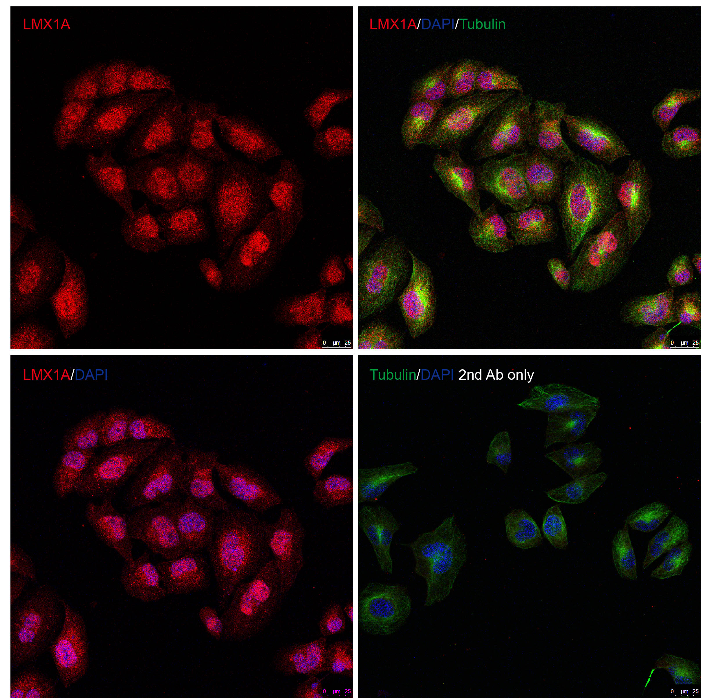
Figure 1 Immunofluorescence Validation of LMX1A in HeLa CellsImmunofluorescent analysis of methanol-fixed HeLa cells labeling LMX1A with 7087 at 10 &956,g/mL, followed by goat anti-rabbit IgG secondary antibody at 1/1000 dilution (red) and DAPI staining (blue). Alpha tubulin was stained with anti-alpha tubulin antibody following by goat anti-mouse IgG secondary antibody (green). Images were captured with confocal microscopy. |
|
|
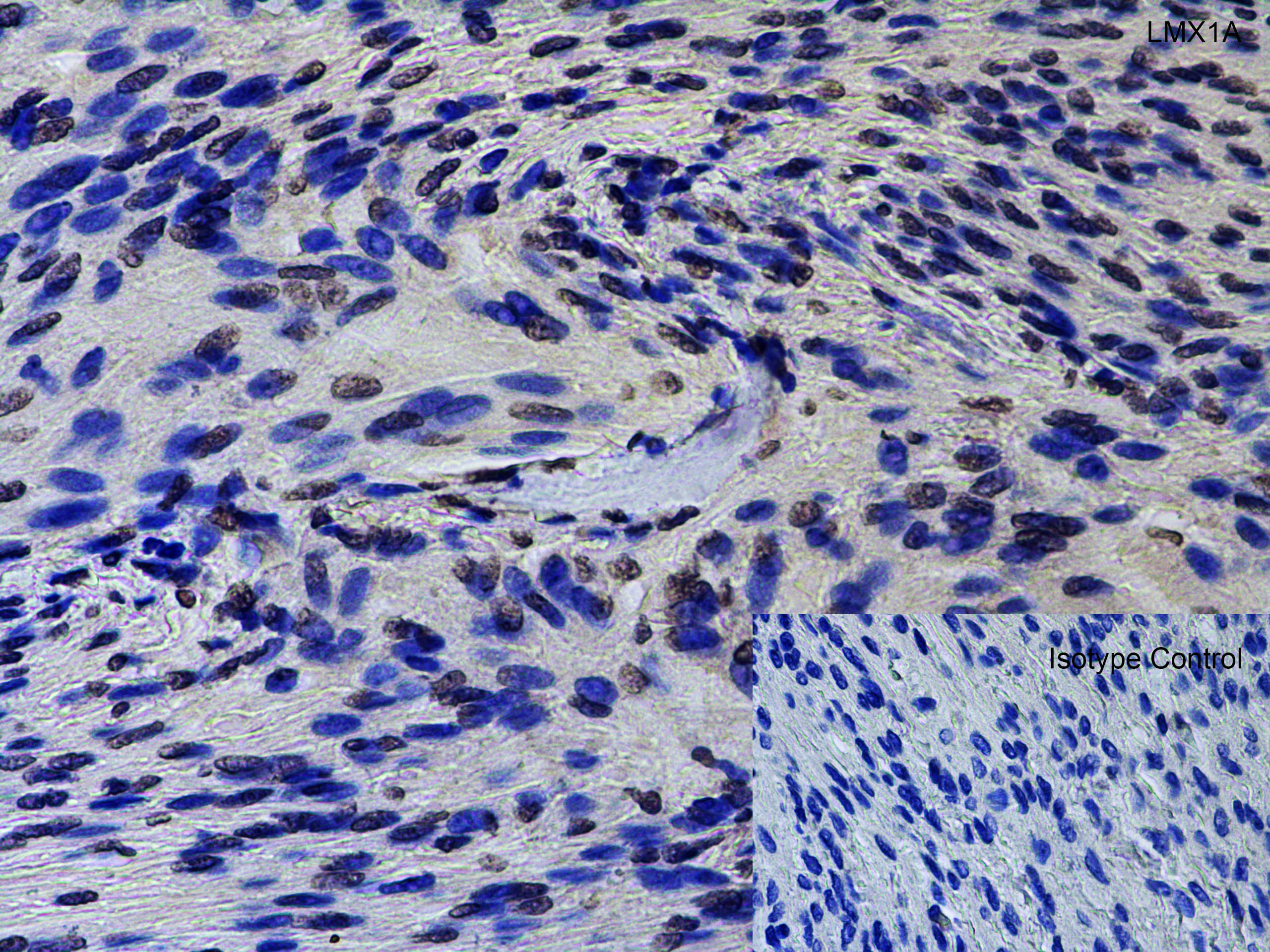
Figure 5 Immunohistochemistry Validation of LMX1A in Human Brain Meningioma Immunohistochemical analysis of paraffin-embedded human brain meningioma using anti-LMX1A antibody (7087) at 2 &956,g/ml. Tissue was fixed with formaldehyde and blocked with 10% serum for 1 h at RT, antigen retrieval was by heat mediation with a citrate buffer (pH6). Samples were incubated with primary antibody overnight at 4730,C. A goat anti-rabbit IgG H&L (HRP) at 1/250 was used as secondary. Counter stained with Hematoxylin. |
|
|
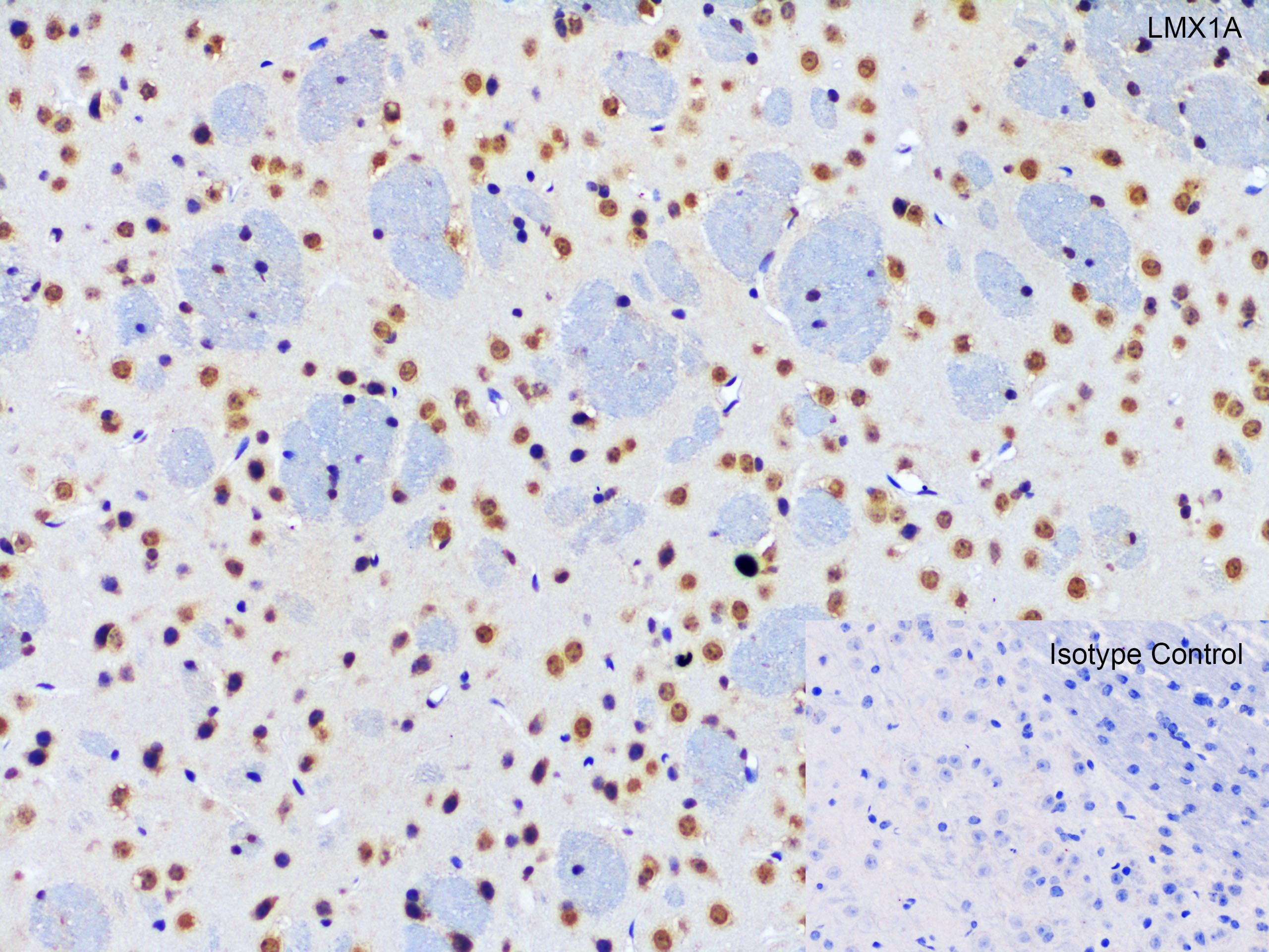
Figure 6 Immunohistochemistry Validation of LMX1A in Mouse Brain Tissue Immunohistochemical analysis of paraffin-embedded mouse brain tissue using anti- anti-LMX1A antibody (7087) at 2 &956,g/ml. Tissue was fixed with formaldehyde and blocked with 10% serum for 1 h at RT, antigen retrieval was by heat mediation with a citrate buffer (pH6). Samples were incubated with primary antibody overnight at 4730,C. A goat anti-rabbit IgG H&L (HRP) at 1/250 was used as secondary. Counter stained with Hematoxylin. |
|
|
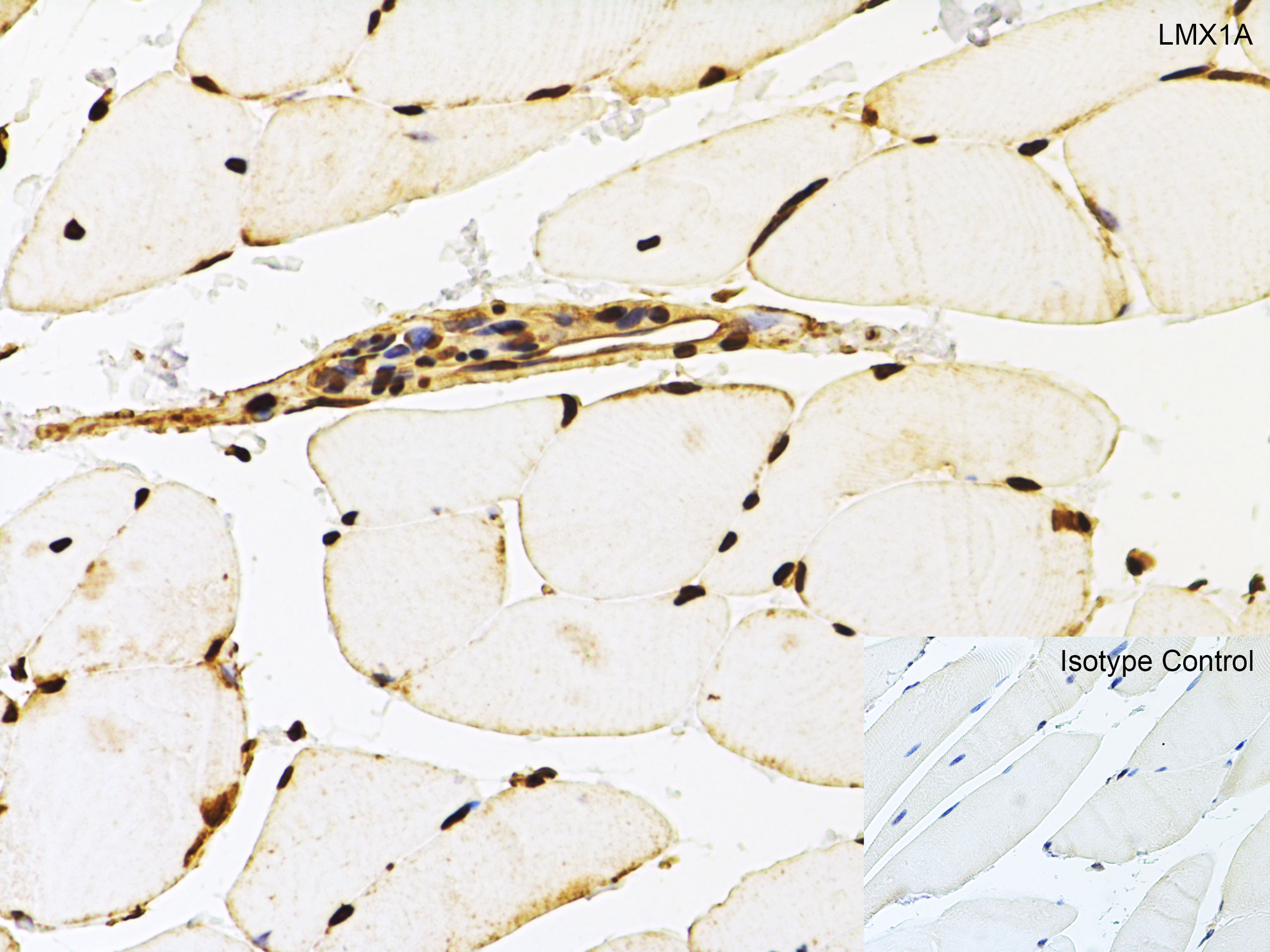
Figure 7 Immunohistochemistry Validation of LMX1A in Rat Skeletal Muscle Tissue Immunohistochemical analysis of paraffin-embedded rat spleen tissue using anti-LMX1A antibody (7087) at 5 &956,g/ml. Tissue was fixed with formaldehyde and blocked with 10% serum for 1 h at RT, antigen retrieval was by heat mediation with a citrate buffer (pH6). Samples were incubated with primary antibody overnight at 4&730,C. A goat anti-rabbit IgG H&L (HRP) at 1/250 was used as secondary. Counter stained with Hematoxylin. |
|
|
Figure 4 Western Blot Validation in Rat Skeletal Muscle Loading: 10 &956,g of lysates per lane.Antibodies: LMX1A, 7087, 1 &956,g/mL, 1h incubation at RT in 5% NFDM/TBST.Secondary: Goat anti-rabbit IgG HRP conjugate at 1:10000 dilution. |
Product Guarantee and Expert Support
