
CRBN Antibody, Unconjugated, Rabbit, Polyclonal
Catalog Number:
PRS-8115
- Images (5)

| Article Name: | CRBN Antibody, Unconjugated, Rabbit, Polyclonal |
| Biozol Catalog Number: | PRS-8115 |
| Supplier Catalog Number: | 8115 |
| Alternative Catalog Number: | PRS-8115-0.02,PRS-8115-0.1 |
| Manufacturer: | ProSci |
| Host: | Rabbit |
| Category: | Antikörper |
| Application: | ELISA, IF, IHC-P, WB |
| Species Reactivity: | Human, Mouse, Rat |
| Immunogen: | CRBN antibody was raised against a 16 amino acid peptide near the carboxy terminus of human CRBN.The immunogen is located within the last 50 amino acids of CRBN. |
| Conjugation: | Unconjugated |
| Alternative Names: | Cereblon, AD-006, DKFZp781K0715, MGC27358, MRT2A |
| Application Dilute: | Optimal dilutions for each application to be determined by the researcher. |
| Application Notes: | CRBN antibody can be used for detection of CRBN by Western blot at 1 - 2 µg/mL. Antibody can also be used for immunohistochemistry starting at 5 µg/mL. For immunofluorescence start at 20 µg/mL.Antibody validated: Western Blot in human samples, Immunohistochemistry in rat samples and Immunofluorescence in rat samples. All other applications and species not yet tested. |
|
|
Figure 2 WB Validation in Human Testis Loading: 15 &956,g of human testis lysate Antibodies: CRBN 8115, 1 h incubation at RT in 5% NFDM/TBST. Secondary: Goat anti-rabbit IgG HRP conjugate at 1:10000 dilution.Lane A: 0.5 &956,g/mLLane B: 1 &956,g/mL |
|
|
Figure 3 WB Validation in Human Cells Loading: 15 &956,g of cell lysate Antibodies: CRBN 8115, 1 &956,g/mL,1 h incubation at RT in 5% NFDM/TBST. Secondary: Goat anti-rabbit IgG HRP conjugate at 1:10000 dilution. |
|
|
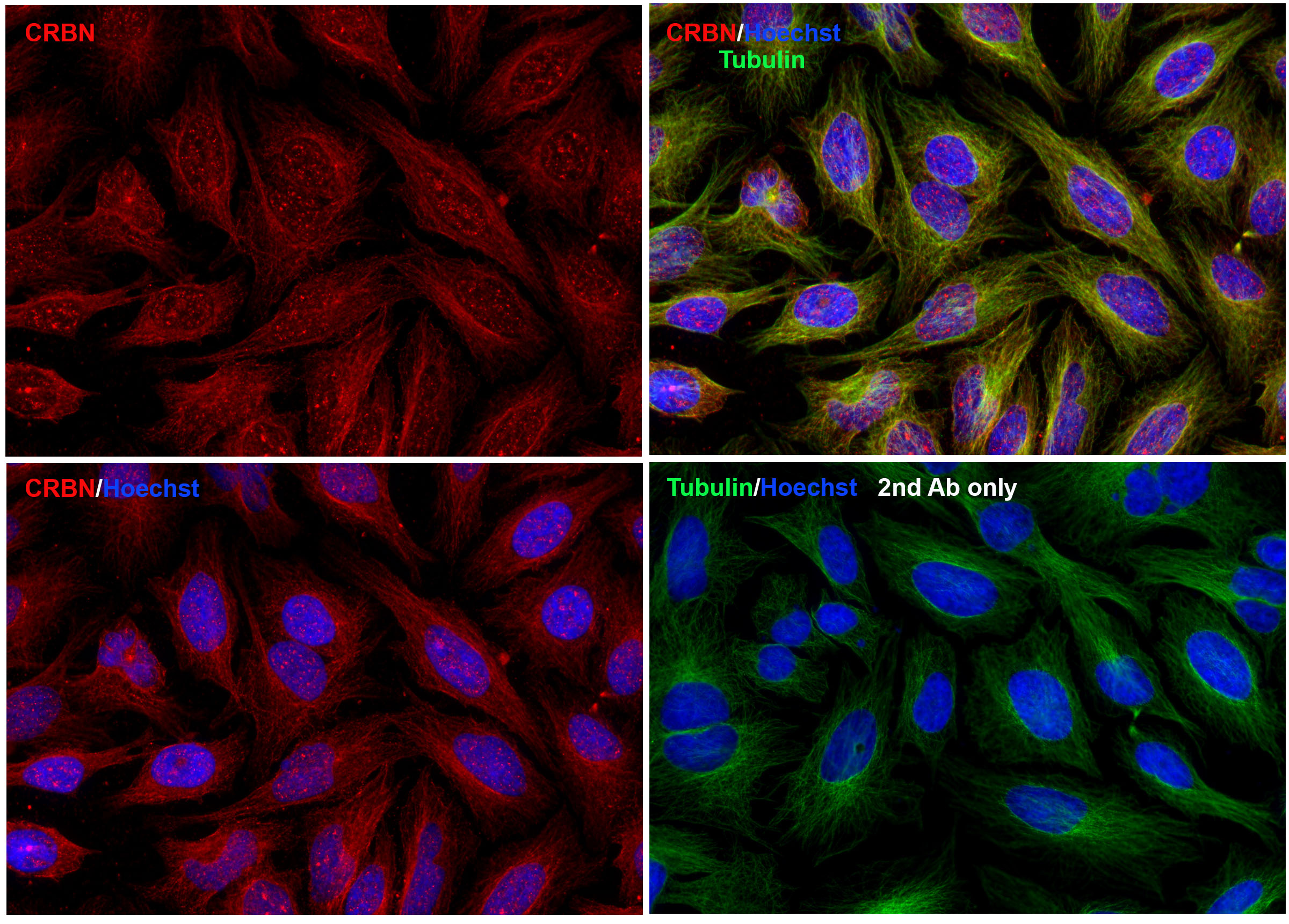
Figure 1 Immunofluorescence Validation of CRBN in HeLa CellsImmunofluorescent analysis of methanol-fixed HeLa cells labeling CRBN with 8115 at 10 &956,g/mL, followed by goat anti-rabbit IgG secondary antibody at 1/1000 dilution (red) and Hoechst staining (blue). Alpha tubulin was stained with anti-alpha tubulin antibody following by goat anti-mouse IgG secondary antibody (green). Images were captured with confocal microscopy. |
|
|
Figure 4 WB Validation in Mouse Tissues Loading: 15 &956,g of mouse tissue lysate Antibodies: CRBN 8115, 0.5 &956,g/mL , 1 h incubation at RT in 5% NFDM/TBST. Secondary: Goat anti-rabbit IgG HRP conjugate at 1:10000 dilution. |
|
|
Figure 5 WB Validation in Rat Tissues Loading: 15 &956,g of rat tissue lysate Antibodies: CRBN 8115, 0.5 &956,g/mL , 1 h incubation at RT in 5% NFDM/TBST. Secondary: Goat anti-rabbit IgG HRP conjugate at 1:10000 dilution. |
Product Guarantee and Expert Support
