
CD80 Detection Set (Risk Free), Unconjugated, Mouse, Monoclonal
Catalog Number:
PRS-RF16040
- Images (9)
| Article Name: | CD80 Detection Set (Risk Free), Unconjugated, Mouse, Monoclonal |
| Biozol Catalog Number: | PRS-RF16040 |
| Supplier Catalog Number: | RF16040 |
| Alternative Catalog Number: | PRS-RF16040-1 |
| Manufacturer: | ProSci |
| Host: | Mouse |
| Category: | Antikörper |
| Application: | ELISA, FC, ICC, IF, IHC-P, WB |
| Species Reactivity: | Human |
| Immunogen: | CD80 antibodies were raised against the extracellular domain of human CD80. |
| Conjugation: | Unconjugated |
| Clonality: | Monoclonal |
| Concentration: | Antibody 1 mg/mL |
| Buffer: | PBS containing 0.02% sodium azide. |
| Form: | Liquid |
| Application Dilute: | Optimal dilutions for each application to be determined by the researcher. |
|
|
Flow cytometry analysis of CD80 overexpressing HEK293 cells using (A) RF16041, (B) RF16042, (C) RF16044, (D) RF16045, (E) RF16046, and (F) control mouse IgG antibody at 10 &956,g/ml. Blue: Untransfected HEK293 cells. Yellow: CD80 overexpressing HEK293 cells. |
|
|
Immunocytochemistry of CD80 in overexpressing HEK293 cells using (A) polyclonal CD80 antibody, (B) RF16041, (C) RF16042, (D) RF16043, (E) RF16044, (F) RF16045, (G) RF16046 and (H) control mouse IgG antibody at 1 &956,g/ml. |
|
|
Immunofluorescence of CD80 in overexpressing HEK293 cells using (A) polyclonal CD80 antibody, (B) RF16041, (C) RF16042, (D) RF16043, (E) RF16044, (F) RF16045, (G) RF16046 and (H) control mouse IgG antibody at 2 &956,g/ml. |
|
|
Immunofluorescence of CD80 in human stomach carcinoma tissue using (A) polyclonal CD80 antibody, (B) RF16041, (C) RF16042, (D) RF16043, (E) RF16044, (F) RF16045, (G) RF16046 and (H) control mouse IgG antibody at 20 &956,g/ml. |
|
|
Immunofluorescence of CD in human tonsil tissue using (A) polyclonal CD80 antibody, (B) RF16041, (C) RF16042, (D) RF16043, (E) RF16044, (F) RF16045, (G) RF16046 and (H) control mouse IgG antibody at 2 &956,g/ml. |
|
|
Immunohistochemistry of CD80 in human stomach carcinoma tissue using (A) polyclonal CD80 antibody at 1 &956,g/ml, (B) RF16041, (C) RF16042, (D) RF16043, (E) RF16044, (F) RF16045, (G) RF16046 and (H) control mouse IgG antibody at 5 &956,g/ml. |
|
|
Immunohistochemistry of CD80 in tonsil tissue using (A) polyclonal CD80 antibody at 1 &956,g/ml, (B) RF16041, (C) RF16042, (D) RF16043, (E) RF16044, (F) RF16045, (G) RF16046 and (H) control mouse IgG antibody at 5 &956,g/ml. |
|
|
Western blot analysis of CD80 in overexpressing HEK293 cells using RF16045 and RF16046 antibody at 0.25 and 0.5 &956,g/ml, respectively. |
|
|
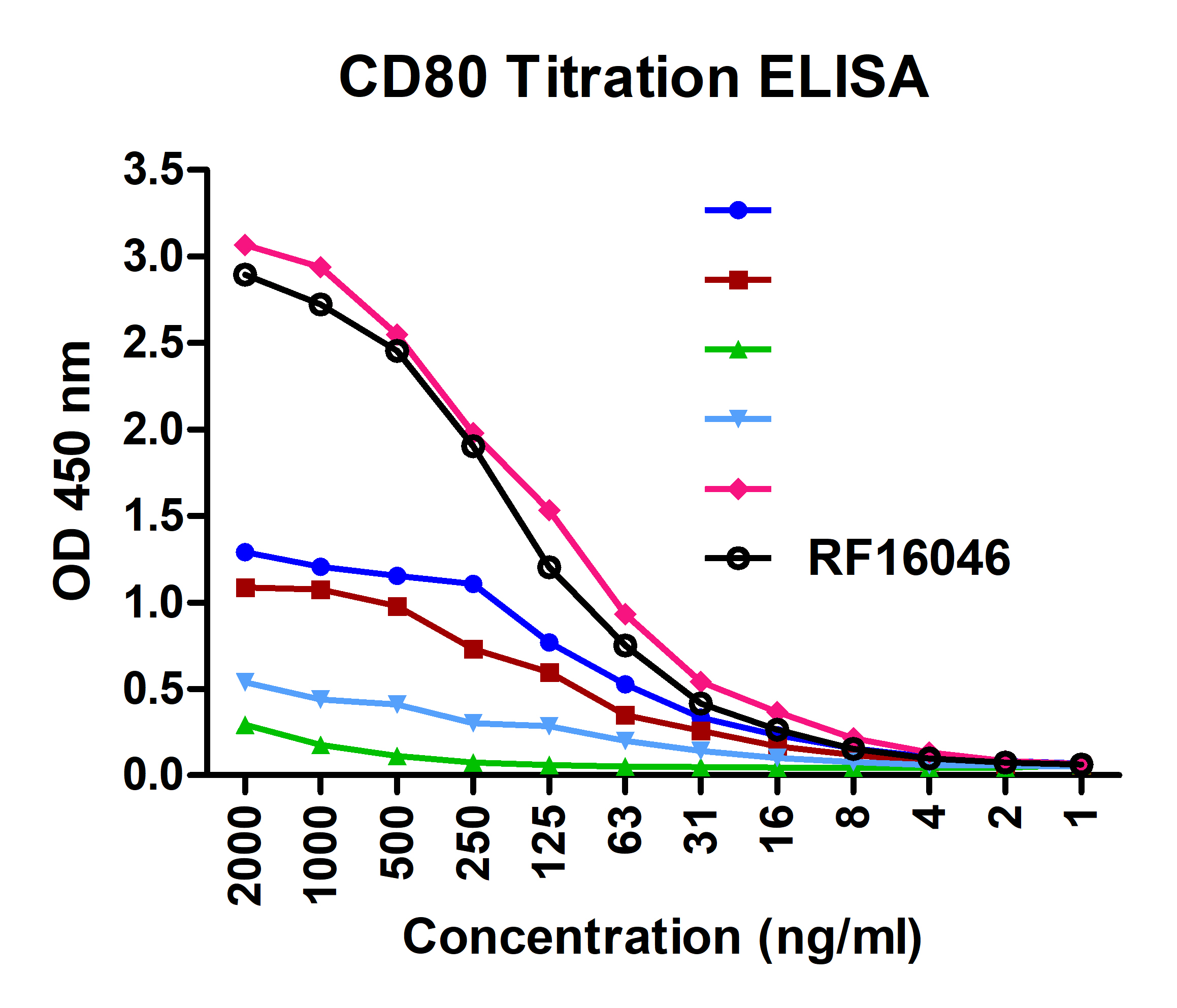
Titration curve analysis of CD80 mAbs to detect recombinant CD80 in ELISA with RF16041, RF16042, RF16043, RF16044, RF16045, and RF16046 abs at decreasing concentrations. |
Product Guarantee and Expert Support
