
Human Collagen Type VI
Catalog Number:
ROC-009-001-108
- Images (8)
| Article Name: | Human Collagen Type VI |
| Biozol Catalog Number: | ROC-009-001-108 |
| Supplier Catalog Number: | 009-001-108 |
| Alternative Catalog Number: | ROC-009-001-108 |
| Manufacturer: | Rockland Immunochemicals |
| Category: | Sonstiges |
| Application: | DOT, SDS-PAGE, WB |
| Conjugation: | Unconjugated |
| Alternative Names: | Type VI collagen, collagen 6, human collagen, collagen VI |
| Application Dilute: | ELISA: 1:10,000, IHC: 1:100-1:500, WB: 1:1000 |
| Application Notes: | Human Collagen type VI purified protein standard has been tested in SDS-Page and is used as a control in ELISA and western blot. Collagen type VI is recognized by type specific anti-collagen antibodies that recognize a native three-dimensional structure. |
|
|
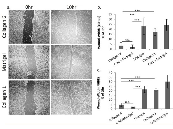
COL6 enhances lung epithelial cell wound repair.(a) Representative images of wound-healing response for 16HBE cells plated on COL6, Matrigel, or COL1. (b-c) Quantitation of wound width at 10 hr post-injury (relative to 0 hr) for 16HBE(b) and NHBE(c) plated on individual matrices, or combinations of matrices. N = 9, ** p<0.01, *** p<0.001. Fig 2. PMID: 30550606. |
|
|
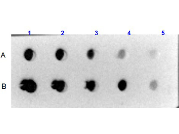
Dot Blot results using Rabbit Anti-Collagen VI Antibody. Sample: A) Human Collagen VI (p/n 009-001-108), B) Bovine/Human Collagen VI (p/n 001-001-108).Load: 1) 1000ng, 2) 333.33ng, 3) 111.11ng, 4) 37.03ng, 5) 12.35ng.Primary Antibody: Rabbit Anti-Collagen VI antibody (p/n 600-401-108) at 5µg/mL at RT for 1hr. Secondary Antibody: Goat Anti-Rabbit IgG HRP Conjugated (p/n 611-1302) 1:40,000 at RT for 30mins. Blocking: BlockOut Universal Blocking Buffer (p/n MB-073) at RT for 30mins. |
|
|
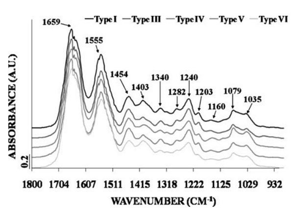
Fourier-transform infrared (FTIR) spectroscopy. Mean FTIR spectra of the five collagen types. Maximal intensity.Fig. 2. PMID: 19685340. |
|
|
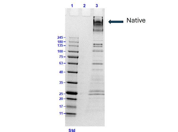
SDS-PAGE results of Human Collagen Type VI. Lane: 1) Opal prestain molecular weight marker (p/n MB-210-0500), 2) blank, 3) Human Collagen Type VI (p/n 009-001-108) [10µg]. Coomassie stained. |
|
|
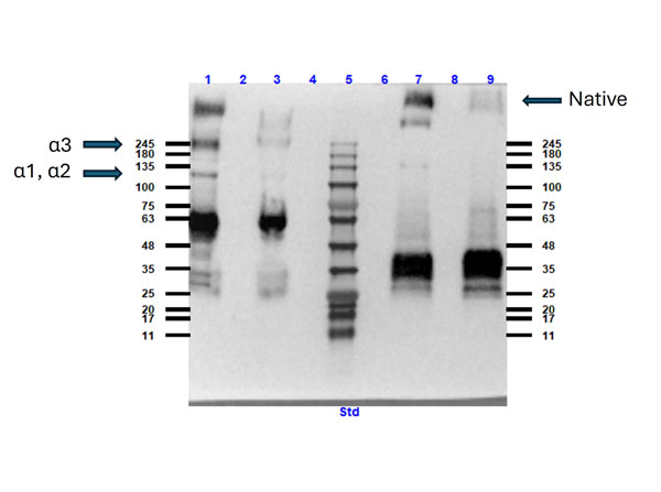
Western blot results using Rabbit Anti-Collagen Type VI Antibody. Lane: 1) Human Collagen Type VI (p/n 009-001-108) reduced and boiled, 3) Human Collagen Type VI reduced and not boiled,5) Opal pre-stained molecular weight marker (p/n MB-210-0500), 7) Human Collagen Type VI not reduced and boiled, 9) Human Collagen Type VI not reduced and not boiled. Lanes 2, 4, 6, 8 blank. Load: 10µg. Primary Antibody: Rabbit Anti-Collagen Type VI Antibody (p/n 600-401-108) 1:500 overnight at 2-8C. Secondary Antibody: Goat anti-Rabbit IgG HRP conjugated (p/n 611-1302) 1:40,000 at RT for 30mins. Blocking: BlockOut Universal Blocking Buffer (p/n MB-073) at RT for 1hr. |
|
|
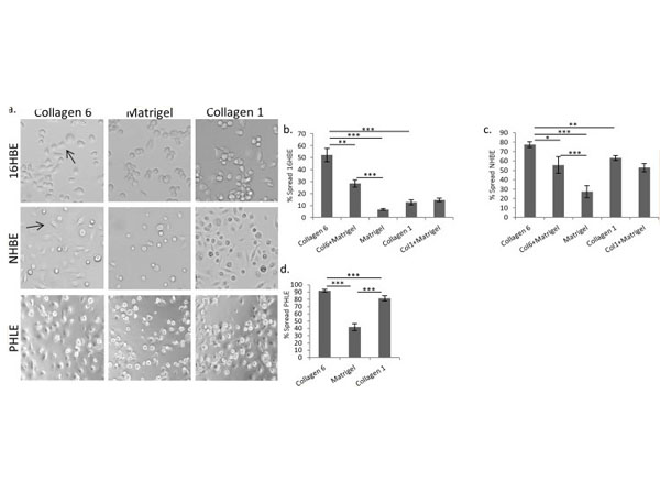
COL6 promotes lung epithelial cell spreading.(a) Representative images of initial, post-adhesion spreading for 16HBE human lung epithelial cells 3 hours after plating cells on COL6, COL1 or Matrigel. Arrows show examples of spread cells. (b-d) Quantitation of the percentage of cells spread 3 hours after plating on different matrices for 16HBE (b), NHBE (c), or PHLE (d) cells. N = 6.* p<0.05, ** p<0.01, *** p<0.001. Fig 3. PMID: 30550606. |
|
|
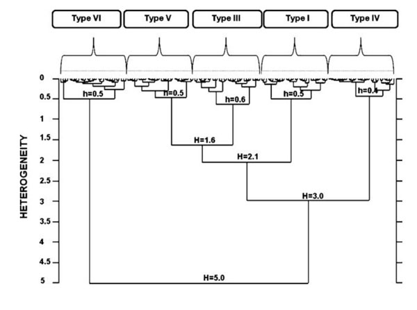
FTIR spectra classification model for the five collagen types. n=30 spectra/collagen type, four spectral intervals, 1,700-1,600 cm-1 , 1,480- 1,350 cm-1 , 1,300-1,180 cm-1 , and 1,100-1,005 cm-1 . h intracluster heterogeneity, H intercluster heterogeneity. Fig. 3. PMID: 19685340. |
|
|
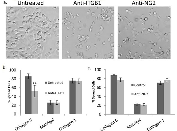
COL6-mediated spreading requires ITGB1, not NG2.(a) Representative images of untreated 16HBE cells, and 16HBE treated with anti-Integrin beta1 or anti-NG2 antibodies 3 hours after plating on COL6. (b) Quantitation of untreated and anti-ITGB1 (b) or anti-NG2 (c) treated 16HBE spreading 3 hours after plating on COL6, COL1, Matrigel coated wells, and uncoated tissue-culture wells (n = 6). * p<0.05, ** p<0.01, *** p<0.001. Fig 4. PMID: 30550606. |
Product Guarantee and Expert Support
