Anti-CIITA antibody has been tested in western blot. For immunoblotting a 1:500 dilution is recommended. Researchers should determine optimal titers for other applications.
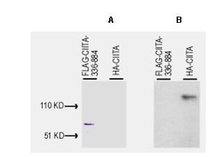
Western blot of Anti-CIITA (1-333) antibody, generated by immunization with bacterially produced FLAG-CIITA aa 1-333, was tested by western blot against lysates of Cos-7 cells after transient transfection, separately, with pcDNA3-FLAG-CIITA-336-884 and pcDNA3-HA-CIITA. For transfection, Fugene 6 (Roche) was used according to the manufacturers instructions for a 6-well plate format. Cells were lysed 24 h post-transfection in 200 µL of 1x SDS-sample buffer, heated at 96C for 5, and vortexed for 30 sec. Samples (10 µL each) were separated on a 12% SDS-PAGE gel and transferred to PVDF (Millipore) followed by blocking for 45 using TTBS supplemented with 5% non-fat dry milk. All incubations were performed at room temperature. In panel A, both samples on PVDF were incubated with 10 µg/mL mouse anti-FLAG antibody (Sigma) for 45. After 5X washes with TTBS, reaction with ALP rabbit anti-mouse IgG at 200 ng/mL proceeded for 45 following again by washing as before. The blot was developed using BCIP/NBT. This blot demonstrates that FLAG-CIITA-336-884 was successfully over-expressed in the Cos-7 cells. In panel B, both samples on PVDF were incubated with a 1:500 dilution of Rocklands anti-CIITA (1-333) for 45. After 5X washes with TTBS, reaction with HRP goat anti-rabbit IgG at 10 ng/mL proceeded for 45 following again by washing as before. The membrane was covered with Pico West Substrate solution (Pierce) for 5 and was then placed between the two layers of a standard sheet protector. Kodak O-MAT film was exposed to the blot for 30 sec and was immediately developed. The lane containing the lysate of pcDNA3-HA-CIITA transfected cells contains a single band at ~130 kDa molecular weight, whereas the lane containing lysate from pcDNA3-FLAG-CIITA-336-884 transfected cells shows no reactivity. This blot demonstrates that anti-CIITA (1-333) is specific for amino acids 1-333 of CIITA and that the antibody is not cross reactive with the FLAG portion of the immunogen.
* VAT and and shipping costs not included. Errors and price changes excepted