Anti-Rabbit IgG HRP, TrueBlot, HRP TrueBlot ULTRA, Peroxidase TrueBlot, TrueBlot for IP/WB, TrueBlot for immunoprecipitation, TrueBlot for western blotting
Clonality:
Monoclonal
Concentration:
1.0 mg/mL by UV absorbance at 280 nm
Clone Designation:
[eB182]
Buffer:
0.02 M Potassium Phosphate, 0.15 M Sodium Chloride, pH 7.2
Form:
Liquid (sterile filtered)
Target:
Rabbit
Application Dilute:
IF Microscopy: User Optimized, WB: 1:1000
Application Notes:
Rabbit IgG HRP TrueBlot has been tested in ELISA, Western blot, and immunoprecipitation and may also be used for detection in immunoblotting assays that do not employ immunoprecipitation. Rabbit IgG TrueBlot is provided as 1000X solution. To conserve re
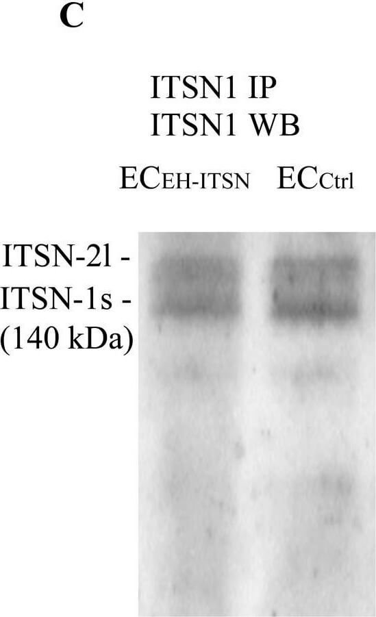
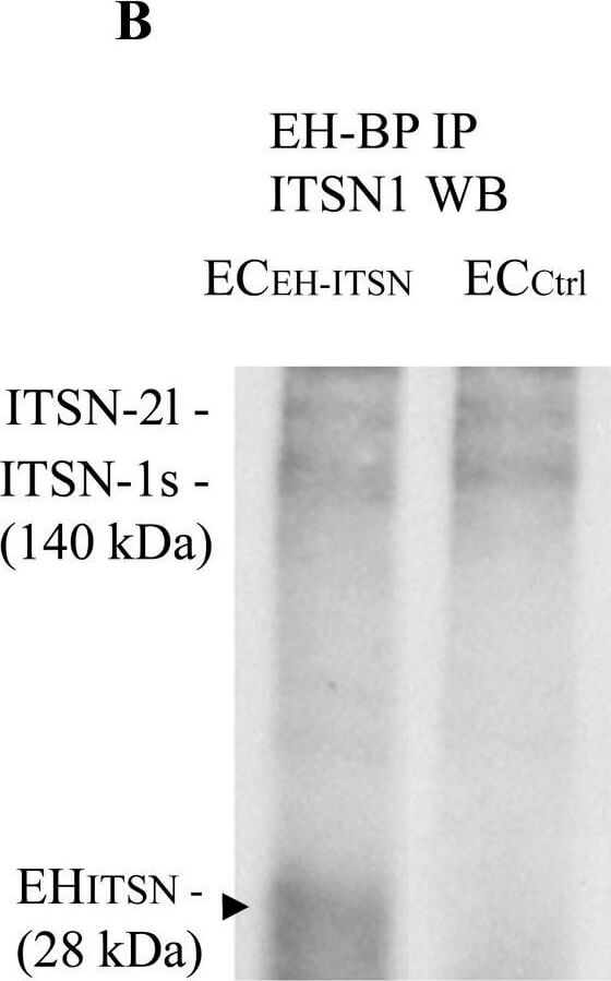
Intersectin-1s (ITSN) interacts via the EH domains with the EHBP1. ECs lysates (250 µg total protein) were subjected to immunoprecipitation with anti-EHBP1 Ab (1 µg), followed by WB with EHBP1 (A) and ITSN1 (B) Abs. EHBP1 Ab brings down the EHBP1 protein as well as ITSN. The 55 kDa immunoreactivity in panel A is cross-reactivity with the IgG heavy chain. The EHBP1 Ab immunoprecipitates the Myc-EHITSN from the stable transfected ECEH-ITSN lysates (B, arrowhead). (C). ECs lysates (250 µg total protein) were subjected to immunoprecipitation with anti-ITSN1 Ab (1 µg), followed by WB with ITSN1 Ab. ITSN1 Ab brings down ITSN protein in both ECEH-ITSN and ECCtrl lysates. The upper ITSN immunoreactivity (190 kDa), belongs to the ITSN-2 long isoform (ITSN-2l). For immunoprecipitation studies (B,C), the rabbit IgG TrueBlot Ab HRP ULTRA conjugated which enables detection o
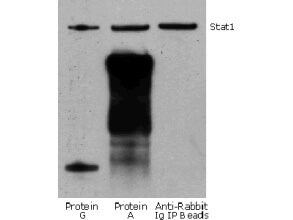
Rabbit TrueBlot IP / Western Blot: Jurkat cell lysate (0.5 ml of 1x10e7 cells/ml) was incubated with rabbit anti-human Stat1 and immunoprecipitated using Protein G, Protein A and Anti-Rabbit Ig IP Beads. Precipitate from 5x10e5 cells was subjected to electrophoresis, transferred to a PVDF membrane, and Western blotted with anti-Stat1 using Rabbit TrueBlot ULTRA: Anti-Rabbit IgG HRP
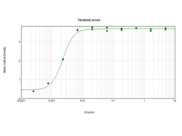
ELISA results of Rabbit TrueBlot ULTRA: Anti-Rabbit IgG HRP tested against purified Rabbit IgG protein. Each well was coated in duplicate with 1.0 µg of Rabbit IgG (p/n 011-0102). The starting dilution of antibody was 5µg/ml and the X-axis represents the Log10 of a 3-fold dilution. The titer is 1:450,000. This titration is a 4-parameter curve fit where the IC50 is defined as the titer of the antibody. Assay performed using 3% fish gelatin as blocking buffer and TMB substrate p/n TMBE-1000.
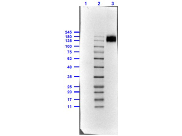
Western Blot of Rabbit TrueBlot ULTRA: Anti-Rabbit IgG HRP. Lane 1: Rabbit IgG WM -reduced (p/n 011-0102) [0.1µg]. Lane 2: Opal Prestained Molecular Weight Marker (p/n MB-210-0500). Lane 3: Rabbit IgG WM non-reduced (p/n 011-0102) [0.1µg]. Antibody: Rabbit TrueBlot ULTRA: Anti-Rabbit IgG HRP at 1.0µg/mL overnight at 4C. Blocking Buffer for Fluorescent Western Blotting (p/n MB-070) for 60mins at RT. Expect: recognizes the Rabbit IgG, only under non-reducing condition. Exposure: 0.45sec.
Intersectin-1s (ITSN) interacts via the EH domains with the EHBP1. ECs lysates (250 µg total protein) were subjected to immunoprecipitation with anti-EHBP1 Ab (1 µg), followed by WB with EHBP1 (A) and ITSN1 (B) Abs. EHBP1 Ab brings down the EHBP1 protein as well as ITSN. The 55 kDa immunoreactivity in panel A is cross-reactivity with the IgG heavy chain. The EHBP1 Ab immunoprecipitates the Myc-EHITSN from the stable transfected ECEH-ITSN lysates (B, arrowhead). (C). ECs lysates (250 µg total protein) were subjected to immunoprecipitation with anti-ITSN1 Ab (1 µg), followed by WB with ITSN1 Ab. ITSN1 Ab brings down ITSN protein in both ECEH-ITSN and ECCtrl lysates. The upper ITSN immunoreactivity (190 kDa), belongs to the ITSN-2 long isoform (ITSN-2l). For immunoprecipitation studies (B,C), the rabbit IgG TrueBlot Ab HRP ULTRA conjugated which enables detection of immunoblotted target protein bands, without interfering with the immunoprecipitating IgG heavy and light chains has been used. (D) Densitometric analysis of immunoprecipitated ITSN in both ECEH-ITSN and ECCtrl lysates. Data are expressed as ratio of ITSN immunoprecipitated by EHBP1 Ab / ITSN immunoprecipitated by ITSN Ab (D). *p < 0.05. (E,F). Double anti-ITSN Ab anti-rabbit IgG Alexa Fluor 594-conjugated (E) / anti-EHBP1 Ab - anti mouse IgG Alexa Fluor 488-conjugated (F). The merged image reveals significant co-localization ITSN/EHBP1, both in the cytosol and at the plasma membrane (G). (H) The magnification of the boxed area in G, highlights the significant co-localization ITSN/EHBP1 at the plasma membrane level (arrows) and cytosol (arrowheads). Bars: 10 µm (E-G), 5 µm (H), n = 5. Figure provided by CiteAb. Source: Front Physiol, PMID: 30333761.
* VAT and and shipping costs not included. Errors and price changes excepted