Anti-Fibrinogen Biotin antibody has been tested by ELISA, western blot, and immunohistochemistry. This product is assayed against 1.0 µg of Fibrinogen in a standard capture ELISA using Peroxidase Conjugated Streptavidin S000-03 and ABTS (2,2-azino-bis-
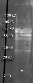
Western Blot of Goat anti-Fibrinogen antibody. Lane 1: Fibrinogen under reducing conditions. Lane 2: none. Load: 1 µg per lane. Primary antibody: Fibrinogen antibody at 1:3000 for overnight at 4C. Secondary antibody: Dylight 488 conjugated Donkey anti goat secondary antibody at 1:10,000 for 45 min at RT. Block: TBS/MB-070 1 hr RT.
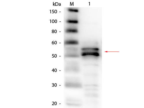
Western Blot of Goat anti-Fibrinogen Antibody (Human Plasma) Biotin Conjugated. Lane 1: Fibrinogen (Human Plasma). Load: 50 ng per lane. Primary antibody: Goat anti-Fibrinogen Antibody (Human Plasma) Biotin Conjugated at 1:1,000 overnight at 4C. Secondary antibody: HRP streptavidin secondary antibody at 1:40,000 for 30 min at RT. Block: MB-070 for 30 minutes at RT. Predicted/Observed size: 56 kDa, 56 kDa for Fibrinogen ß-chain.
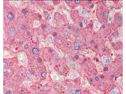
Immunohistochemistry of Goat Anti-Fibrinogen antibody. Tissue: human liver tissue. Fixation: formalin fixed paraffin embedded. Antigen retrieval: not required. Primary antibody: Fibrinogen antibody at 1:500 for 1 h at RT. Secondary antibody: Peroxidase goat secondary antibody at 1:10,000 for 45 min at RT. Localization: Fibrinogen is localized in plasma. Staining: Fibrinogen as precipitated red signal with hematoxylin purple nuclear counterstain.
* VAT and and shipping costs not included. Errors and price changes excepted