Anti-AKT pS473 antibody was produced by repeated immunizations with a synthetic peptide corresponding to residues surrounding S473 of human AKT1 protein, followed by hybridoma development
0.02 M Potassium Phosphate, 0.15 M Sodium Chloride, pH 7.2
Form:
Liquid (sterile filtered)
Target:
Human
Antibody Type:
Primary Antibody
Application Dilute:
ELISA: 1:20,000, Flow Cytometry: User Optimized, IHC: 20 µg/mL, IF Microscopy: 1:500 - 1:3,000, IP: User Optimized, WB: 1:500 - 1:3,000
Application Notes:
This monoclonal antibody is tested in ELISA, immunofluorescence, immunohistochemistry, flow cytometry, and western blotting. Expect a band approximately 56 kDa in size corresponding to phosphorylated AKT protein by western blotting in the appropriate cel
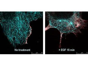
Immunofluorescence confocal microscopy of Mouse Anti-AKT pS473 antibody. Tissue: EGF treated A431 cells. Fixation: 0.5% PFA. Antigen retrieval: EGF 15 min. Primary antibody: AKT pS473 antibody at 10 µg/mL for 1 h at RT. Secondary antibody: DyLight 488(TM) Goat anti-Rabbit IgG, MAb anti-AKT pS473, atto-647N anti-Mouse IgG (Active Motif). at 1:10,000 for 45 min at RT. Localization: AKT pS473 is nuclear and occasionally cytoplasmic. Staining: AKT pS473 as red signal with tubulin (cyan).
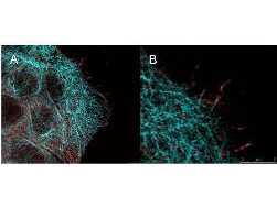
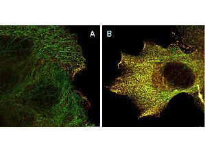
High resolution STED immunofluorescence nanoscopy of Mouse anti-AKT pS473 antibody. Tissue: A431 cells. The merge images (A) and at high magnification (B) show phosphorylated AKT colocalized with the distal microtubules. Fixation: 4% paraformaldehyde for 5 min and after washes blocked with 10% NGS/0.2% Triton X-100 for 30 min. Antigen retrieval: serum deprivation for 12 h. Primary antibody: AKT pS473 antibody at 10 µg/mL and alpha-tubulin (cyan) (p/n 600-401-880) at 1.4 µg/mL for 1 h at RT. Secondary antibody: Atto 647N anti-Mouse IgG (ATTO TEC GmbH), and DyLight(TM)488 anti-Rabbit IgG (p/n 611-141-122) were used at 1.0 µg/mL for 1h at RT for indirect detection. Localization: AKT pS473 is in the cytoplasm and also organized at the periphery of the cell. Staining: AKT pS473 as red signal with bis-benzimide (blue) nuclear counterstain.
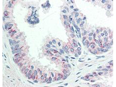
Immunohistochemistry of Mouse anti-AKT pS473 antibody. Tissue: human prostate tissue. Fixation: formalin fixed paraffin embedded. Antigen retrieval: not required. Primary antibody: AKT pS473 antibody at 20 µg/mL for 1 h at RT. Secondary antibody: Dakos Techmate streptavidin-biotin reagents at 1:10,000 for 45 min at RT. Localization: AKT pS473 is nuclear and occasionally cytoplasmic. Staining: AKT pS473 as precipitated red signal with hematoxylin purple nuclear counterstain.
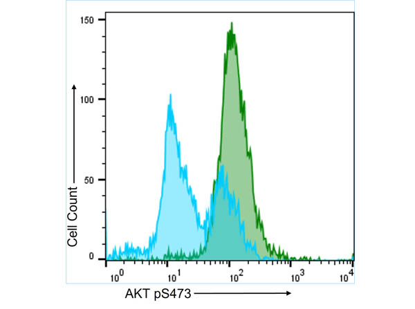
Flow Cytometry results of Anti-AKT pS473 (MOUSE) Monoclonal Antibody. The green histogram represents the A431 cells that were stimulated for 15 minutes with 100 ng/mL EGF. The blue histogram shows the untreated A431 cell population, which is bimodal. Both populations were stained with a 1:50 dilution of the Anti-AKT pS473 (MOUSE) Monoclonal Antibody (p/n 200-301-268) for 30 mins at 4C. The secondary antibody, Anti-MOUSE IgG (H&L) (GOAT) Antibody DyLight(TM) 488 Conjugated (p/n 610-141-002) was used at a 1:200 dilution for 30 mins at 4C.
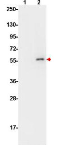
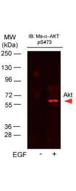
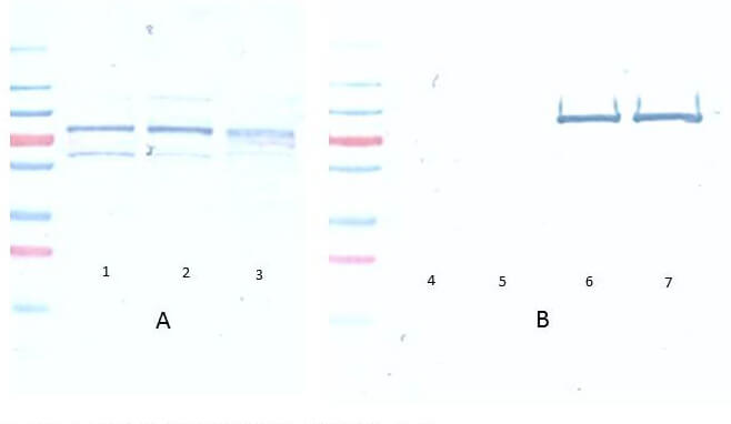
Western Blot of Mouse Anti-AKT pS473 antibody. Lane 1: non-phosphorylated AKT in untreated cells. Lane 2: phosphorylated AKT (indicated by arrowhead at ~56 kDa) on PDGF stimulated NIH/3T3 cell lysates. Load: 10 µg per lane. Primary antibody: AKT pS473 antibody at 1:10,000 in TBS with 0.05% Tween-20 with 1% BSA, for 1 h at 4 C. Secondary antibody: HRP conjugated Gt-a-Mouse IgG (p/n 610-103-121) was used at a 1:20,000 dilution for 1 h at 4 C with FemtoMax(TM) enhanced chemiluminescent reagent (p/n FEMTOMAX-100). Other band(s): none.
Immunofluorescence Microscopy of Mouse Anti-AKTpS473 antibody using STED nanoscopy to evaluate AKT activation and migration. Tissue: A431 cells. Antigen retrieval: Panel A: serum starved,unstimulated cells. Panel B: serum starved, EGF stimulated for 15 mins. A massive increase in AKT-pS473 activation, as measured by intensity signal, peaked at 15 minutes and was associated with depolymerized tubulin. Staining: Panel A shows STED data (AKT-pS473, red channel) collected simultaneously with confocal signal (a-tubulin, green channel). Upon stimulation of cells with EGF, a rapid activation of AKT is observed (Panel B) along with a coincident change in the tubulin organization (yellow signal), as well as an extensive cell shape-change (cell membrane folding) and accumulation of AKTpS473 at the cell periphery.
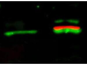
Western Blot of Mouse Anti-Akt pS473 antibody. Lane 1: unstimulated NIH/3T3 lysates contain inactive unphosphorylated Akt1, green band. Lane 2: PDGF stimulated NIH/3T3 lysate contains both inactive (green band) and activated ph
* VAT and and shipping costs not included. Errors and price changes excepted