
RFP Antibody, IgG2a, Clone: [8E5.G7], Mouse, Monoclonal
Catalog Number:
ROC-200-301-379
- Images (13)

| Article Name: | RFP Antibody, IgG2a, Clone: [8E5.G7], Mouse, Monoclonal |
| Biozol Catalog Number: | ROC-200-301-379 |
| Supplier Catalog Number: | 200-301-379 |
| Alternative Catalog Number: | ROC-200-301-379 |
| Manufacturer: | Rockland Immunochemicals |
| Host: | Mouse |
| Category: | Antikörper |
| Application: | ELISA, WB |
| Species Reactivity: | Other |
| Immunogen: | The immunogen is a Red Fluorescent Protein (RFP) fusion protein corresponding to the full-length amino acid sequence (234aa) derived from the mushroom anemone Discosoma. |
| Conjugation: | Unconjugated |
| Alternative Names: | mouse anti-RFP Antibody, DsRed, rDsRed, Discosoma sp. Red Fluorescent Protein, Red fluorescent protein drFP583 |
| Application Dilute: | ELISA: 1:75,000 - 1:150,000, Flow Cytometry: User Optimized, IF Microscopy: User Optimized, IP: User Optimized, WB: 1:1,000 - 1:10,000 |
| Application Notes: | Monoclonal anti-RFP is designed to detect RFP and its variants. This antibody has been tested by ELISA and Western blot, and is suitable for use in FISH, IF, IHC, and multiplex assays based on published references. This antibody can be used to detect RFP |
|
|
|
|
|
|
|
|
|
|
|
|
|
|
|
|
|
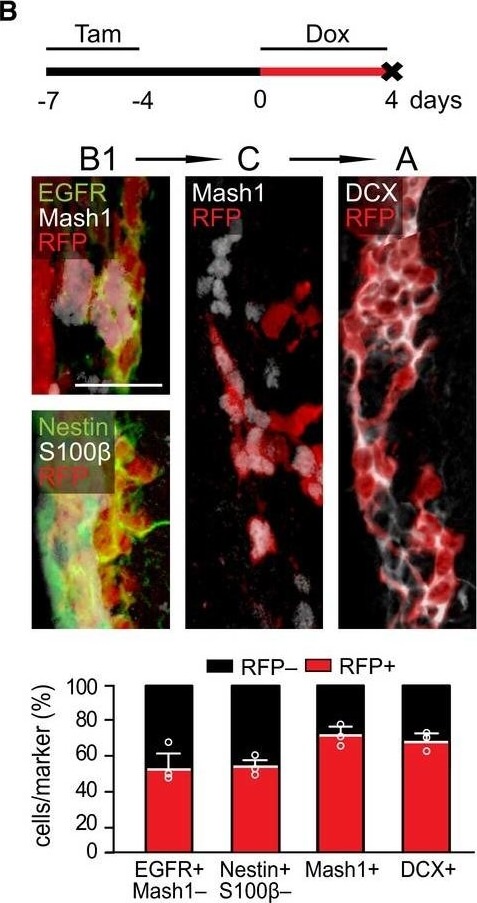
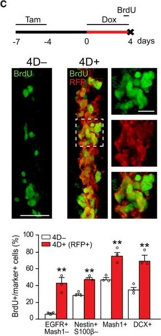
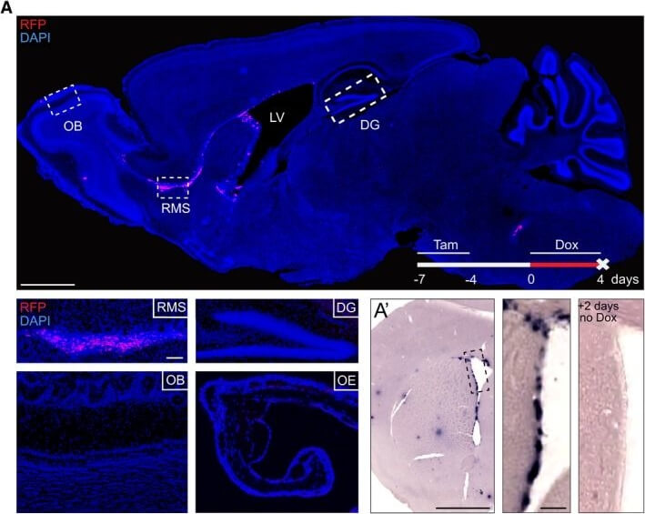
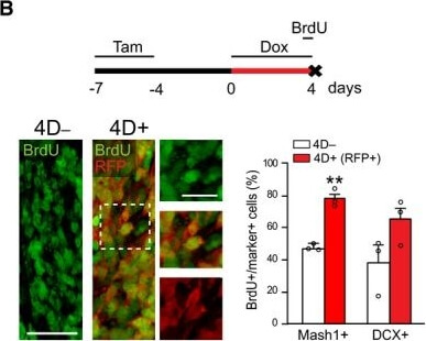
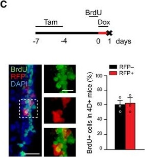
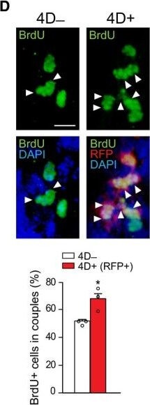
Characterization of the transgenic model and effect of 4D on the RMSA Fluorescence image of a sagittal section of a 4D+ brain after a 4-day treatment with doxycycline showing RFP signal confined to the SVZ and RMS (nuclei counterstained with DAPI, blue). Insets show representative images of specific brain regions (as indicated) and the olfactory epithelium. APhase contrast picture of the SVZ upon insitu hybridization against mRNA for RFP in a 4D+ brain treated as in (A) and sacrificed immediately after (left) or 2days after (right) doxycycline administration. (B, C) Experimental design (top), fluorescence pictures (left with magnified insets), and quantifications (right) of BrdU incorporation in the RMS (B) or SVZ (C). (B) shows the proportion of BrdU in C (Mash1+) and A (DCX+) cells in 4D- (white) and 4D+ (red, among RFP+) mice. (C) shows the proportion of RFP- (black) and RFP+ (red) among BrdU+ cells of 4D+ mice. (A) OB, olfactory bulb, RMS, rostral migratory stream, LV, lateral ventricle, DG, dentate gyrus, OE, olfactory epithelium. (A-C) Tam, tamoxifen, Dox, doxycycline. (B, C) MeanSEM, **P<0.01, unpaired Students t-test, N=3 mice and n>1,100 cells. Scale bars=500 (A top, A), 100 (insets A and A), 50 (B and C), and 20 (insets B and C) µm. Figure provided by CiteAb. Source: EMBO J, PMID: 30643018. |
|
|
|
|
|
|
|
|
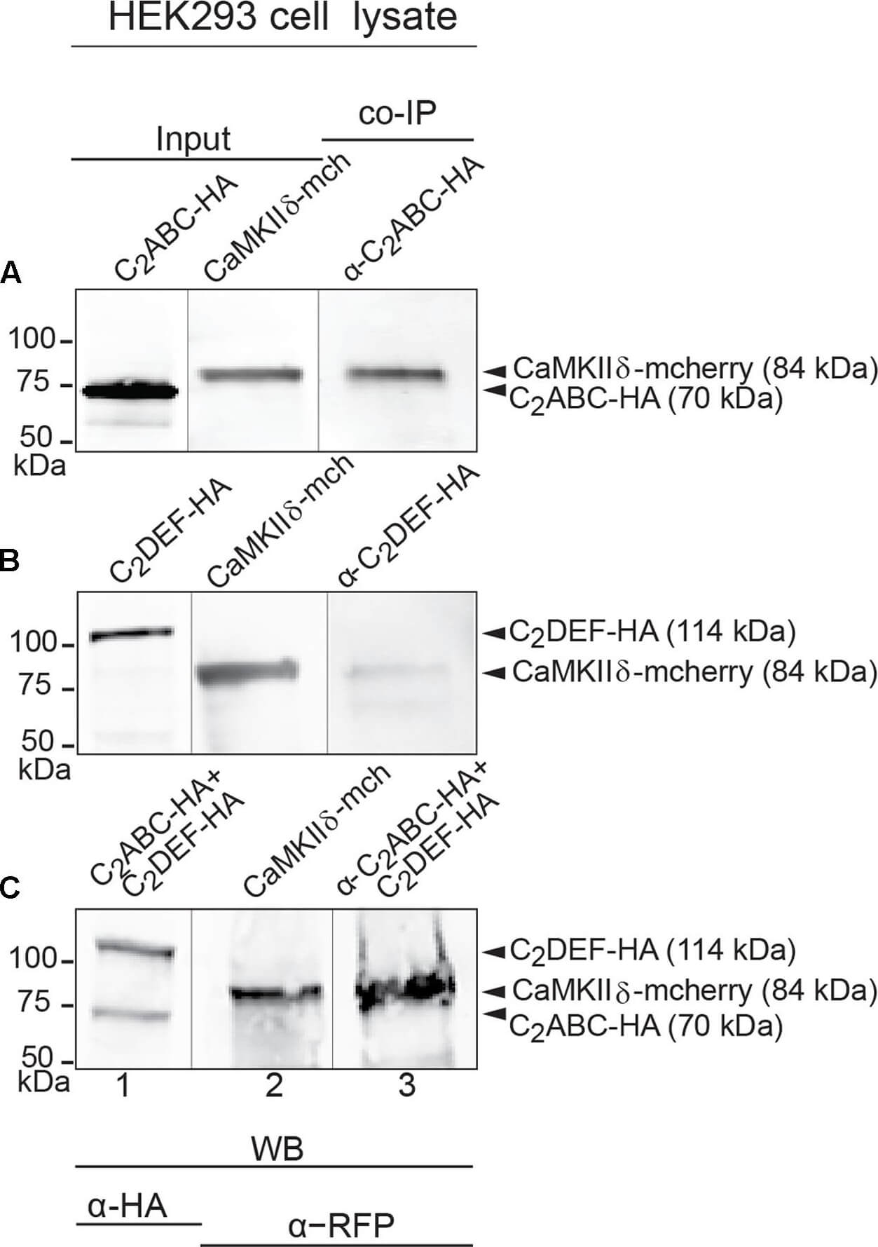
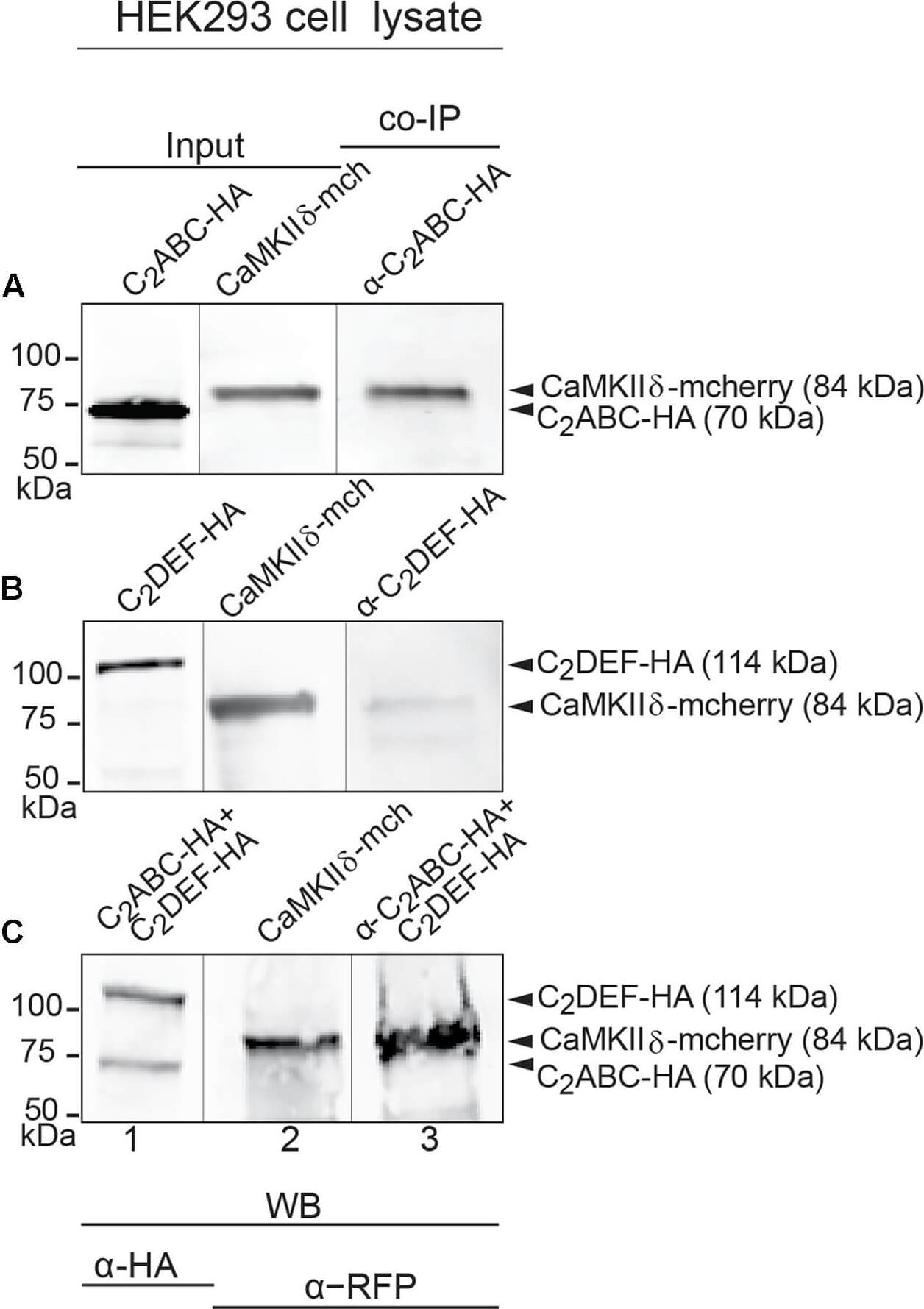
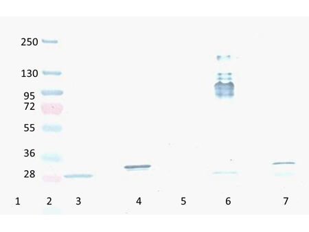
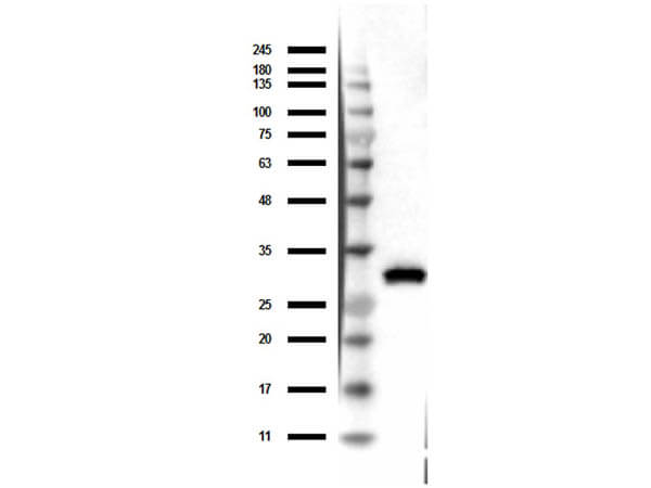
Western Blot of Mouse Anti-RFP Antibody. Lane 1: Opal Prestain Molecular weight (p/n MB-210-0500). Lane 2: 50ng of RFP. Primary Antibody: Mouse Anti-RFP at 1µg/mL overnight at 2-8C. Secondary Antibody: Rabbit Anti-Mouse HRP (p/n 610-403-C46) at 1:40,000 for 30mins at RT. Block: BlockOut Universal blocking buffer (p/n MB-073). Expect ~27kDa. |
|
|
Characterization of the transgenic model and effect of 4D on the RMSA Fluorescence image of a sagittal section of a 4D+ brain after a 4-day treatment with doxycycline showing RFP signal confined to the SVZ and RMS (nuclei counterstained with DAPI, blue). Insets show representative images of specific brain regions (as indicated) and the olfactory epithelium. APhase contrast picture of the SVZ upon insitu hybridization against mRNA for RFP in a 4D+ brain treated as in (A) and sacrificed immediately after (left) or 2days after (right) doxycycline administration. (B, C) Experimental design (top), fluorescence pictures (left with magnified insets), and quantifications (right) of BrdU incorporation in the RMS (B) or SVZ (C). (B) shows the proportion of BrdU in C (Mash1+) and A (DCX+) cells in 4D- (white) and 4D+ (red, among RFP+) mice. (C) shows the proportion of RFP- (black) and RFP+ (red) among BrdU+ cells of 4D+ mice. (A) OB, olfactory bulb, RMS, rostral |
|
|
|
|
|
Product Guarantee and Expert Support
