This Protein A purified antibody was prepared by repeated immunizations with a synthetic peptide corresponding to a region near the carboxy terminal end of human NAG-1 protein. A residue of cysteine was added to facilitate coupling to KLH.
0.02 M Potassium Phosphate, 0.15 M Sodium Chloride, pH 7.2
Form:
Liquid (sterile filtered)
Target:
Human
Antibody Type:
Primary Antibody
Application Dilute:
ELISA: 1:2,000, WB: 1:1,000
Application Notes:
This Protein A purified Anti-NAG1 antibody has been tested by ELISA and western blotting for human NAG-1 protein. For detection of NAG-1 in human serum, a sandwich ELISA is suggested using this antibody in combination with anti-NAG-1/GDF15 (N-terminal),
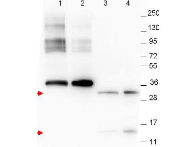
Western blot using Rocklands anti-NAG-1 monoclonal antibody. The blot shows detection of recombinant NAG-1 protein present in Pichia pastoris whole cell lysates: lane 1 - yeast cell lysate expressing NAG-1 H variant with SUMO expression tag at 36 kDa, lane 2 - yeast cell lysate expressing NAG-1 D variant with SUMO expression tag at 36 kDa, lane 3 - yeast cell lysate expressing NAG-1 H variant, and lane 4 - yeast cell lysate expressing NAG-1 D variant. Recombinant NAG-1 proteins without SUMO correspond to monomer (15 kDa) and dimer (30 kDa) bands as indicated by the arrowheads. All lysates were run under reducing conditions. Primary antibody was used at a 1:1,000 dilution in TBS contains 1% BSA and 0.2% Tween, and reacted overnight at 4C. For detection, a 1:40,000 dilution of peroxidase conjugated Gt-a-Mouse IgG secondary antibody (610-103-121) was used in Blocking Buffer for Fluorescent Western Blotting (MB-070) for 30 min at room temperature. Molecular weight estimation was made by comparison to prestained MW markers. Image was captured using the BioRad Versadoc(TM) 4000MP Imaging System.
* VAT and and shipping costs not included. Errors and price changes excepted