This monoclonal antibody was produced by repeated immunizations with a synthetic peptide corresponding to an internal region of human LGR4 protein. The hybridoma was produced by fusing BALB/c mouse splenocytes and mouse myeloma SP2/O cells using conventional technology.
Anti-LGR4 monoclonal antibody has been tested by ELISA and western blot, and is suitable in immunohistochemistry. Expect a band approximately 102 kDa in size corresponding to LGR4 protein by western blotting in the appropriate cell lysate or extract. Spe
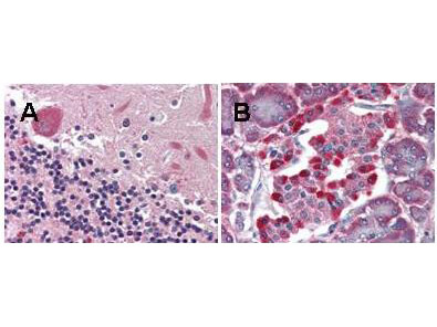
Rocklands anti-LGR4 monoclonal antibody was used diluted to 5 µg/ml to detect LGR4 staining at the membrane of cells in various human tissues. A. Brain cerebellum. B. Pancreas islet. Strongly positive staining is noted in subsets of cells within the islets of Langerhans. Moderately positive staining was observed in Purkinje and Golgi neurons of the cerebellum, adrenal medulla, neuroendocrine cells, hepatocytes, lung macrophages, seminiferous tubules and Leydig cells of the testis. Faintly to moderately positive staining was also observed in cardiac myocytes and renal tubules, granulocytes, and subsets of lymphocytes. Some elastin background staining is noted. Tissue was formalin fixed and paraffin embedded. No pre-treatment of sample was required. The image shows the localization of antibody as the precipitated red signal, with a hematoxylin purple nuclear counterstain. Personal communication, Andrew Elston, Lifespan Biosciences, Seattle, WA.
* VAT and and shipping costs not included. Errors and price changes excepted