Cav3.2 Antibody was produced in mice by repeated immunizations with a fusion protein corresponding to an internal region (II-III loop) of human Cav3.2.
Conjugation:
Unconjugated
Alternative Names:
CACNA1HB, Alpha1 3.2, CACNA 1H, CACNA1 HB, Cav T.2, EIG6, Voltage dependent T type calcium channel alpha 1H, Low-voltage-activated calcium channel alpha1 3.2 subunit, Voltage-gated calcium channel subunit alpha Cav3.2
0.02 M Potassium Phosphate, 0.15 M Sodium Chloride, pH 7.2
Form:
Liquid (sterile filtered)
Target:
Human
Antibody Type:
Primary Antibody
Application Dilute:
IHC: 0.1-1.0ug/mL, IF Microscopy: 1.0-10ug/mL, IP: User Optimized, WB: 1-10ug/mL
Application Notes:
Anti-CAV3.2 Antibody is tested by WB, IP, IHC, and IF microscopy. Expect a band approximately ~260kDa on specific lysates. Specific conditions for reactivity should be optimized by the end user.
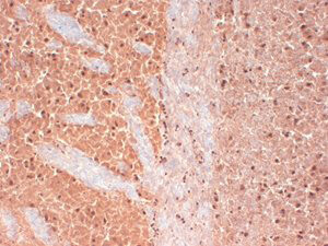
Immunohistochemistry of Mouse anti-Cav3.2 antibody. Tissue: mouse brain extract. Fixation: Frozen. Primary Antibody: anti-Cav 3.2 antibody at 1ug/ml for 1h at RT. Secondary antibody: Peroxidase mouse secondary at 1:10,000 for 45 min at RT. Localization: Membrane. Staining: Cav 3.2 as precipitated brown signal.
* VAT and and shipping costs not included. Errors and price changes excepted