Anti-AKT pT308 monoclonal antibody was produced by repeated immunizations with a synthetic peptide corresponding to residues surrounding T308 of human AKT1 protein.
0.02 M Potassium Phosphate, 0.15 M Sodium Chloride, pH 7.2
Form:
Lyophilized
Target:
Human
Antibody Type:
Primary Antibody
Application Dilute:
ELISA: 1:20,000, Flow Cytometry: User Optimized, IHC: 20 µg/ml, IP: User Optimized, WB: 1:500 - 1:3,000
Application Notes:
Biotin Conjugated Anti-AKT pT308 is tested for ELISA, immunohistochemistry, immunoprecipitation and western blotting. Expect a band approximately 56 kDa in size corresponding to phosphorylated AKT protein by western blotting in the appropriate cell lysat
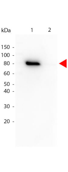
Western Blot of Mouse anti-Akt phospho T308 Biotin Conjugated antibody. Lane 1: GST tagged AKT1 active recombinant protein. Lane 2: GST tagged AKT1 un-active recombinant protein. Load: 25 ng per lane. Primary antibody: Akt phospho T308 Biotin Conjugated antibody at 1:1,000 for overnight at 4C. Secondary antibody: HRP Streptavidin secondary antibody at 1:40,000 for 30 min at RT. Block: MB-070 for 30 min at RT. Predicted/Observed size: 79 kDa, 79 kDa for Akt phospho T308. Other band(s): none
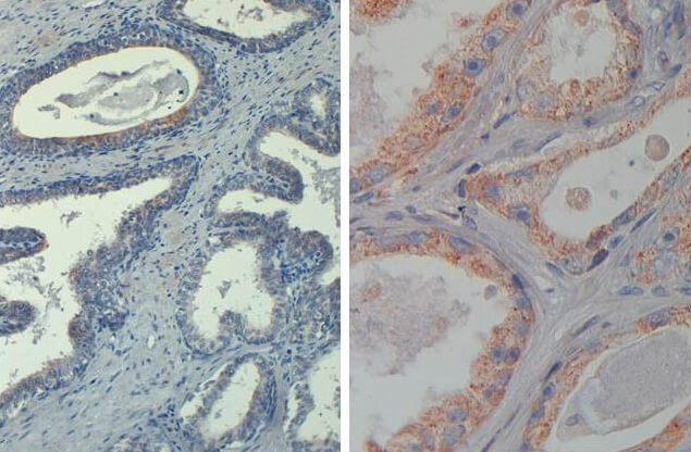
Immunohistochemistry of Mouse Anti-AKT phospho T308 Biotin Conjugated. Tissue: prostate at 20X (left) and 40X (right). Fixation: FFPE buffered formalin 10% conc. Antigen retrieval: Heat, Citrate pH 6.2. Pressure Cooker , Heat, EDTA pH 9.5 Pressure Cooker. Primary antibody: AKT pT308 biotin at 20 µg/mL for 1 h at RT. Secondary antibody: Streptavidin Conj. HRP at 10 ug/ml. Localization: nuclear and occasionally cytoplasmic. Staining: antibody as precipitated red signal with a hematoxylin purple nuclear counterstain.