AKT3 PE Antibody, IgG1, Clone: [25F6.F6.D8], RPE, Mouse, Monoclonal
Biozol Catalog Number:
ROC-200-308-E75
Supplier Catalog Number:
200-308-E75
Alternative Catalog Number:
ROC-200-308-E75
Manufacturer:
Rockland Immunochemicals
Host:
Mouse
Category:
Antikörper
Application:
DOT, ELISA
Species Reactivity:
Human, Mouse, Rat
Immunogen:
Anti-AKT3 Antibody was prepared from tissue culture supernatant by Protein A affinity chromatography using a synthetic peptide corresponding to internal residues of human AKT3 protein.
Conjugation:
RPE
Alternative Names:
Mouse anti-AKT3 antibody PE conjugation, phycoerythrin conjugated Mouse anti-AKT 3 antibody, AKT-3, PKB antibody, PKB gamma antibody, PKBGAMMA antibody, PRKBG antibody, Protein kinase Akt 3 antibody, Protein kinase B gamma antibody, RAC-gamma serine/threonine-protein kinase, RAC-PK-gamma
0.02 M Potassium Phosphate, 0.5 M Sodium Chloride, pH 7.2
Form:
Lyophilized
Target:
Human
Antibody Type:
Primary Antibody
Application Dilute:
ELISA: User Optimized, Flow Cytometry: User Optimized, IHC: User Optimized, IF Microscopy: User Optimized, WB: User Optimized
Application Notes:
Anti-AKT3 PE Antibody has been tested by ELISA and dot blot and is suitable for Flow Cytometry, immunohistochemistry, and western blotting. Expect a band approximately 56 kDa in size corresponding to AKT3 protein by western blotting in the appropriate ce
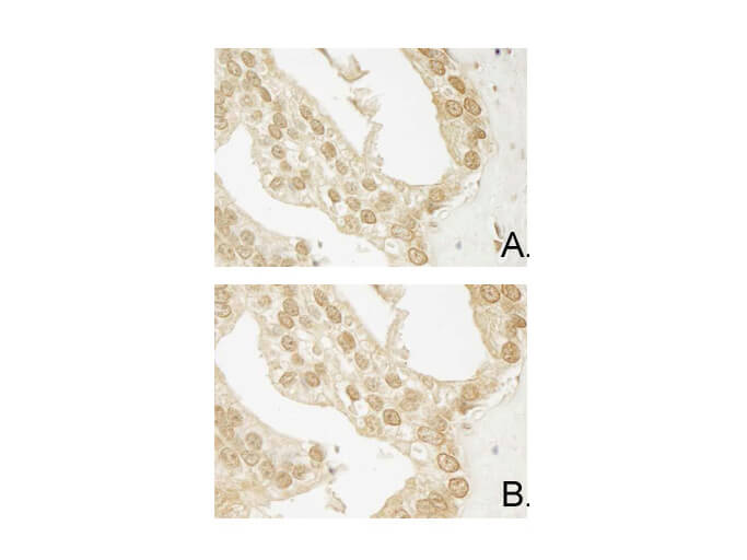
Immunohistochemistry of Mouse Anti-AKT3 antibody. Tissue: human prostate carcinoma. A) AKT-3 antibody produced using CELLine, B) AKT-3 antibody produced using roller bottle. Fixation: formalin fixed paraffin embedded. Antigen retrieval: not required. Primary antibody: AKT-3 antibody at 10 µg/mL for 1 h at RT. Secondary antibody: Peroxidase mouse secondary antibody at 1:10,000 for 1 h at RT. Localization: AKT3 is nuclear and occasionally cytoplasmic. Staining: AKT3 as precipitated brown signal with hematoxylin purple nuclear counterstain.
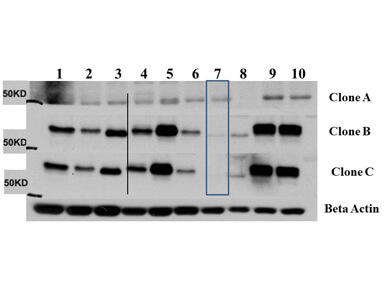
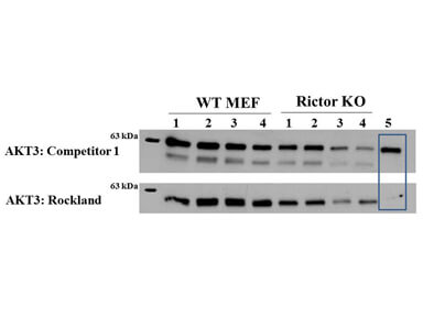
Western Blot of Mouse Anti-AKT3 antibody. Lane 1: C2C12. Lane 2: MEF1. Lane 3: MEF2. Lane 4: A549. Lane 5: Calu-1. Lane 6: PC3. Lane 7: HepG2. Lane 8: Jurkat. Lane 9: SKOV3. Lane 10: 293T. Load: 35 µg per lane. Primary antibody: AKT-3 antibody at 1:1000 for overnight at 4C. Secondary antibody: Anti mouse secondary antibody at 1:20,000 for 1 h at RT. Block: 5% BLOTTO overnight at 4C. Predicted/Observed size: 56 kDa for AKT3.
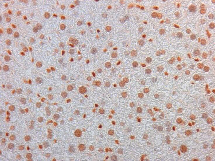
Immunohistochemistry of Mouse Monoclonal anti AKT3 Antibody in Mouse Embryonic Kidney. Tissue: Mouse Liver. Fixation: FFPE buffered formalin 10% conc. Ag Retrieval: Heat, Citrate pH 6.2. Pressure Cooker. Primary antibody: anti-AKT3 at 2ug/ml for 1.5 hour room Temp. Secondary Ab: MOUSE ON MOUSE HRP POLYMER 45 RT.
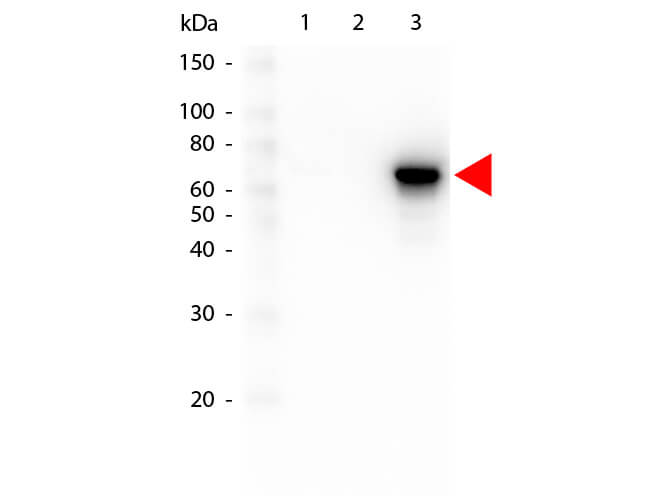
Western Blot of Mouse anti-AKT3 antibody. Lane 1: GST Tagged recombinant AKT1. Lane 2: GST Tagged recombinant AKT2. Lane 3: GST Tagged recombinant AKT3. Load: 25 ng per lane. Primary antibody: AKT3 antibody at 1:1,000 for overnight at 4C. Secondary antibody: Peroxidase mouse secondary antibody at 1:40,000 for 30 min at RT. Block: MB-070 for 30 min at RT. Predicted/Observed size: 78 kDa for AKT3. Other band(s): none.
Western Blot of Mouse anti-AKT3 antibody. Lane 1: Control. Lane 2: Rapa. Lane 3: T50. Lane 4: T250. Lane 5: Control. Lane 6: Rapa. Lane 7: T50. Lane 8: T250. Lane 9: AKT3 null. Load: 35 µg per lane. Primary antibody: AKT-3 antibody at 1:1000 for overnight at 4C. Secondary antibody: Anti mouse secondary antibody at 1:20,000 for 1 h at RT. Block: 5% BLOTTO overnight at 4C. Predicted/Observed size: 56 kDa for AKT3.
* VAT and and shipping costs not included. Errors and price changes excepted