Anti-CXCR4-Lo antibody was prepared from whole rabbit serum produced by repeated immunizations with a peptide corresponding to nine amino acids near the N-terminus of human CXCR4 isoform a.
0.01 M Sodium Phosphate, 0.25 M Sodium Chloride, pH 7.2
Form:
Liquid (sterile filtered)
Target:
Human
Antibody Type:
Primary Antibody
Application Dilute:
ELISA: 1:100,000, IF Microscopy: 4 µg/mL, WB: 10 µg/mL
Application Notes:
Anti-CXCR4-Lo Antibody has been tested for use in ELISA, Western Blotting, Immunocytochemistry, and Immunofluorescence. Specific conditions for reactivity should be optimized by the end user. Expect a band at approximately 40 kDa in Western Blots of spec
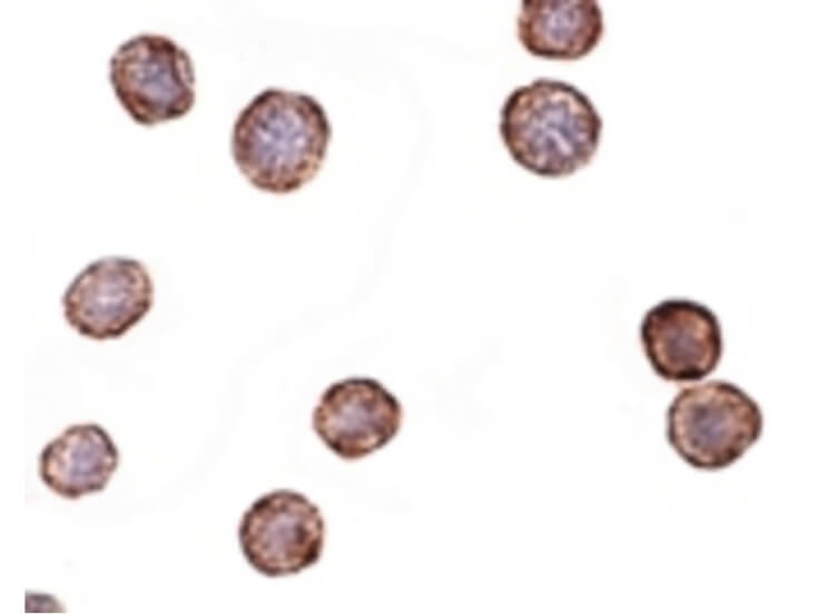
Immunohistochemistry of CXCR4-Lo antibody. Tissue: HeLa cells. Fixation: formalin fixed paraffin embedded. Antigen retrieval: not required. Primary antibody: CXCR4-Lo antibody at 2 µg/mL for 1 h at RT. Secondary antibody: Peroxidase rabbit secondary antibody at 1:10,000 for 45 min at RT. Localization: CXCR4-Lo is nuclear and occasionally cytoplasmic. Staining: CXCR4-Lo as a precipitated red signal with hematoxylin purple nuclear counterstain.
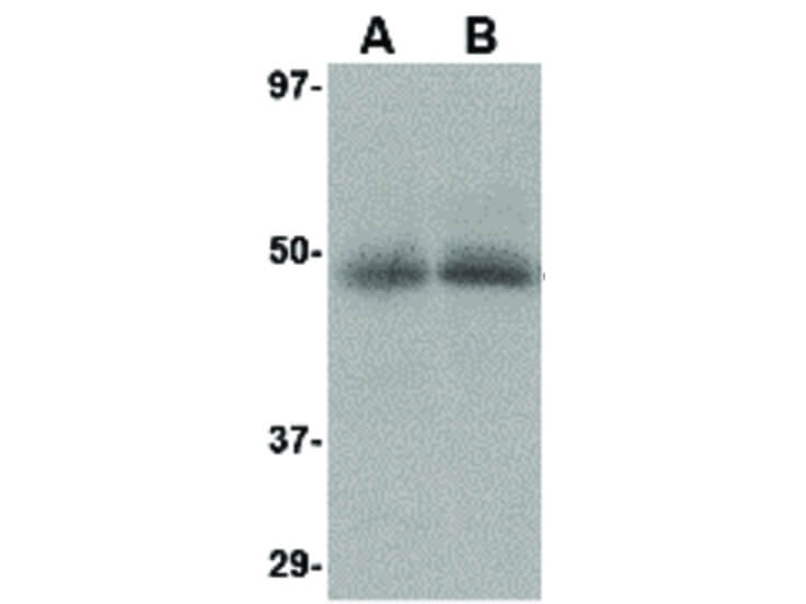
Western Blot of CXCR4-Lo antibody. Lane A: Human spleen tissue lysate at 10 µg/mL. Lane B: Human thymus tissue lysate at 10 µg/mL. Load: 35 µg per lane. Secondary antibody: Peroxidase rabbit secondary antibody at 1:10,000 for 45 min at RT. Block: 5% BLOTTO overnight at 4C. Predicted/Observed size: 39.7 kDa, 49 kDa for CXCR4-Lo.
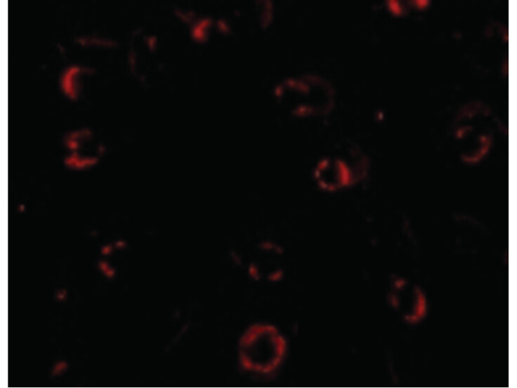
Immunofluorescence Microscopy of CXCR4-Lo antibody. Tissue: HeLa cells. Fixation: 0.5% PFA. Antigen retrieval: not required. Primary antibody: CXCR4-Lo antibody at 4 µg/mL for 1 h at RT. Secondary antibody: Fluorescein rabbit secondary antibody at 1:10,000 for 45 min at RT. Staining: CXCR4-Lo as a red fluorescent signal.
* VAT and and shipping costs not included. Errors and price changes excepted