CXCR4 Antibody was produced from whole rabbit serum prepared by repeated immunizations with a peptide corresponding to amino acids near the n-terminus of human CXCR4.
Anti-CXCR4 Antibody is tested for use in E, WB, ICC, IP, IF, and FACS. Expect a band approximately ~39.7 kDa on specific lysates. Western Blot in human, mouse, and rat samples, Immunohistochemistry, Immunocytochemistry and Immunofluorescence in human sam
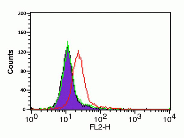
Flow Cytometry Validation of CXCR4Cells: HeLa cells. Overlay histogram showing HeLa cells stained with Anti-CXCR4 at 1µg/1x106 cells (red line) for 1h at 4C in 2% FBS/PBS. Followed by secondary antibody 488 goat anti-rabbit IgG (H+L) at 1:500 dilution for 1h at 4C. Isotype control mouse IgG1 antibody at 1µg/1x106 cells (green line) used under the same conditions.
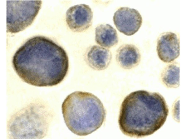
Immunocytochemistry Validation of CXCR4. Cells: HeLa cells. Fixation: formaldehyde and blocked with 10% serum for 1 h at RT. Antigen retrieval: heat mediation with a citrate buffer (pH6). Primary: anti-CXCR4 antibody at 2 µg/ml overnight at 4C. Secondary: goat anti-rabbit IgG H&L (HRP) at 1:250 was used as secondary. Counter stained with Hematoxylin.
Immunofluorescence Validation of CXCR4. Cells: HeLa cells. Fixation: 4% paraformaldehyde. Primary: Anti-CXCR4 at 20 µg/mL. Secondary: goat anti-rabbit IgG at 1:500 dilution (red). Image showing both membrane and cytoplasmic staining on HeLa cells.
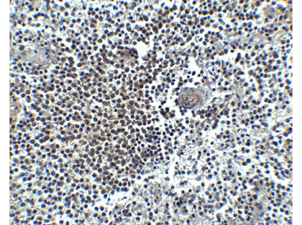
Immunohistochemistry Validation of CXCR4. Tissue: Human Spleen. Fixation: paraffin-embedded, formaldehyde and blocked with 10% serum for 1 h at RT. Antigen retrieval: heat mediation with a citrate buffer (pH6). Primary: anti-CXCR4 antibody at 5 µg/ml overnight at 4C. Secondary: goat anti-rabbit IgG H&L (HRP) at 1:250. Counter stained with Hematoxylin.
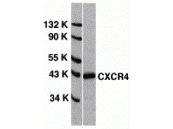
Western Blot Validation of CXCR4.Loading: 15 µg of HeLa cell lysates per lane. Primary Antibody: Anti-CXCR4 at 1µg/mL for 1h at RT in 5% NFDM/TBST. Secondary: Goat anti-rabbit IgG HRP conjugate at 1:10000 dilution.
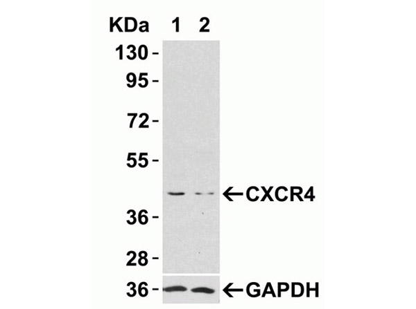
Western Blot Validation of CXCR4 siRNA Knockdown. Loading: 10 µg of HeLa whole cell lysates per lane. HeLa cells were transfected with control siRNAs (lane 1) or CXCR4 siRNAs (lane 2). Primary Antibody: Anti-CXCR4 at 2µg/mL for 1h at RT in 5% NFDM/TBST. Secondary: Goat anti-rabbit IgG HRP conjugate at 1:10000 dilution.
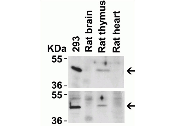
Animal Species Reactivity Western Blot Validation of CXCR4. Loading: 20 µg per lane. Lane 1: 293 lysate, Lane 2: Rat Brain lysate, Lane 3: Rat Thymus lysate, Lane 4: Rat Heart lysate. Primary Antibody: top panel Anti-CXCR4 (p/n 200-401-G91) at 2µg/mL, bottom panel (p/n 600-401-G92) at 2µg/mL for 1h at RT in 5% NFDM/TBST. Secondary: Goat anti-rabbit IgG HRP conjugate at 1:10000 dilution.
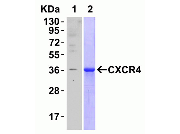
Recombinant Protein Test Western Blot Validation of CXCR4. Loading: CXCR4 partial recombinant protein (Novus Biologicals, Cat H00007852-Q01). Lane 1: Primary antibody Anti-CXCR4 antibody at 0.1 µg/mL for 1h at RT in 5% NFDM/TBST. Secondary: Goat anti-rabbit IgG HRP conjugate at 1:10000 dilution. Lane 2: Coomassie blue staining.
* VAT and and shipping costs not included. Errors and price changes excepted