DR5 Antibody was produced from whole rabbit serum prepared by repeated immunizations with a peptide corresponding to amino acids near the c-terminus of human DR5.
0.02 M Potassium Phosphate, 0.15 M Sodium Chloride, pH 7.2
Form:
Liquid
Target:
Human
Antibody Type:
Primary Antibody
Application Dilute:
Flow Cytometry: User Optimized, IHC: User Optimized, IF Microscopy: User Optimized, WB: 5 µg/mL
Application Notes:
Anti-DR5 Antibody is tested for use in WB, Flow, Flow-CS, ICC/IF, IHC, IHC-P. Expect a band approximately 47.9 kDa on specific lysates. Specific conditions for reactivity should be optimized by the end user.
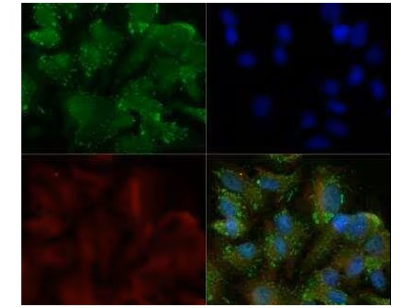
Immunocytochemistry/Immunofluorescence analysis of Anti-DR5/TRAIL R2/TNFRSF10B Antibody. HepG2 cells were fixed for 10 minutes using 10% formalin and then permeabilized for 5 minutes using 1X TBS + 0.5% Triton-X100. The cells were incubated with anti-DR5 at 5ug/ml overnight at 4C and detected with an anti-rabbit Dylight 488 (Green) at a 1:500 dilution. Alpha tubulin (DM1A) was used as a co-stain at a 1:1000 dilution and detected with an anti-mouse Dylight 550 (Red) at a 1:500 dilution. Nuclei were counterstained with DAPI (Blue). Cells were imaged using a 40X objective.
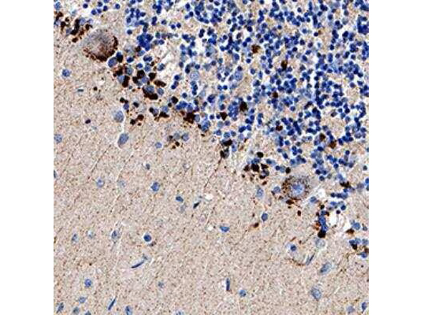
Immunohistochemistry analysis of Anti-DR5/TRAIL R2/TNFRSF10B antibody at 1:200 concentration, formalin fixed paraffin-embedded (FFPE) human brain cerebellum, on a Bond Rx autostainer (Leica Biosystems). The assay involved 30 minutes of heat induced antigen retrieval (HIER) using 10mM sodium citrate buffer (pH 9.0) and endogenous peroxidase quenching with peroxide block. The sections were incubated with primary antibody for 15 minutes and Bond Polymer Refine Detection (Leica Biosystems) with DAB was used for signal development followed by counterstaining with hematoxylin. Cytoplasmic staining was observed in the Purkinje cell layer.
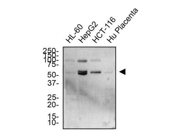
Western Blot analysis of Anti-DR5/TRAILR2/TNFRSF10B Antibody. Total protein from HL-60, HepG2, HCT-116 and human placenta was separated on a 12% gel by SDS-PAGE, transferred to PVDF membrane and blocked in 5% non-fat milk in TBST. The membrane was probed with 2.0 ug/ml anti-DR5 in 1% non-fat milk in TBST and detected with an anti-rabbit HRP secondary antibody using chemiluminescence.
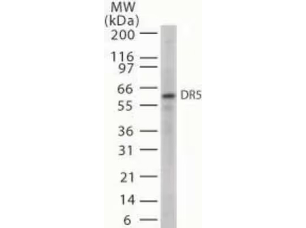
Western Blot analysis of Anti-DR5/TRAILR2/TNFRSF10B Antibody. Detection of 20 ug of whole cell lysates from HL60 cells with anti-D5 at 5µg/ml.
* VAT and and shipping costs not included. Errors and price changes excepted