0.02 M Potassium Phosphate, 0.15 M Sodium Chloride, pH 7.2
Form:
Liquid (sterile filtered)
Target:
Human
Antibody Type:
Primary Antibody
Application Dilute:
ELISA: 1:200, Gel Shift: User Optimized, IF Microscopy: 10ug/ml, IP: User Optimized, WB: 1 µg/mL
Application Notes:
Anti-HSF1 Antibody is tested for WB, IP, IF microscopy, and IHC. Expect a band approximately ~85kDa protein in unstressed cell lysates, and an ~95kDa protein in heat shocked cell lysates, corresponding to the molecular mass of inactive and active forms o
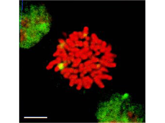
Immunofluorescence of rat anti-HSF1 antibody. Tissue: mitotic HeLa cells. Antigen retrieval: Heat Shock. Primary Antibody: HSF1 antibody at 10ug/ml for 1h at RT. Secondary antibody: Fluorescein rat secondary at 1:10,000 for 45 min at RT. Localization: nuclear. Staining: HSF1 granules present as (green) fluorescent signal.
* VAT and and shipping costs not included. Errors and price changes excepted