0.02 M Potassium Phosphate, 0.15 M Sodium Chloride, pH 7.2
Form:
Liquid (sterile filtered)
Target:
Human
Antibody Type:
Primary Antibody
Application Dilute:
ELISA: 1:1,000 - 1:5,000, IHC: 1:100 - 1:500, IF Microscopy: User Optimized, WB: 1:500 - 1:2,000
Application Notes:
This IgG fraction antibody of anti-Human TNFalpha has been tested for use in neutralizations, ELISA, immunohistochemistry and immunoblotting. It recognizes the 17,000 MW TNFalpha. Reactivity in other immunoassays is unknown.
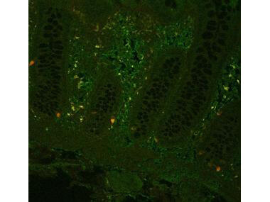
Fluorescent immunohistochemistry showing staining of human colon by Rocklands anti-TNF alpha (formalin/PFA-fixed paraffin-embedded sections). Samples were formaldehyde-fixed, then blocked in 10% serum for 2 hours at 20C. The primary antibody was diluted 1:100 and incubated with the sample for 2 hours at 20C. Alexa Fluor 680 goat polyclonal secondary antibody was used diluted 1:5000.
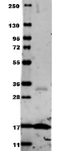
Western blot using Rocklands Anti-Human TNF-a (RABBIT) Antibody. Membrane blocked in 1% BSA-TBS-T for 30 min at RT, Rb-a-TNF alpha added at 1:1000 in 1% BSA-TBS-T o/n 4C, DyLight 649 Gt-a-Rb (p/n 611-143-122) added at 1:20,000 in buffer (p/n MB-070) for 30 min at RT.
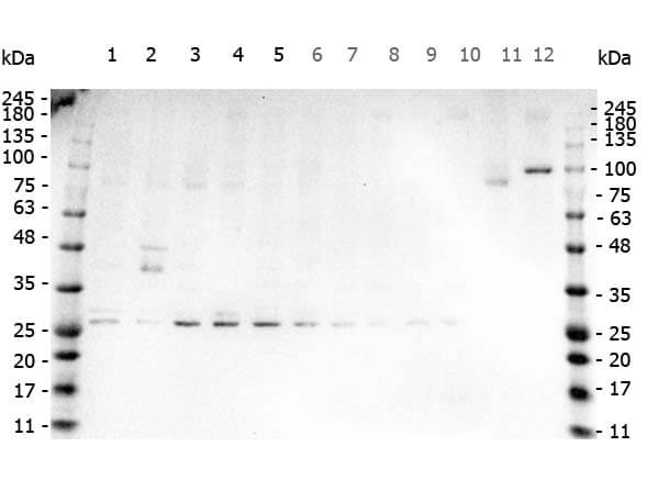
Western Blot of Rabbit anti-TNF Alpha antibody. Marker: Opal Pre-stained ladder (p/n MB-210-0500). Lane 1: HEK293 lysate (p/n W09-000-365). Lane 2: HeLa Lysate (p/n W09-000-364). Lane 3: MCF-7 Lysate (p/n W09-000-360). Lane 4: Jurkat Lysate (p/n W09-000-370). Lane 5: A431 Lysate (p/n W09-000-361). Lane 6: A549 Lysate (p/n W09-001-372). Lane 7: LNCap Lysate (p/n W09-001-GJ9). Lane 8: MOLT-4 Lysate (p/n W09-001-GK2). Lane 9: Ramos Lysate (p/n W09-000-GK4). Lane 10: Raji Lysate (p/n W09-001-368). Lane 11: A-172 Lysate (p/n W09-001-GL5). Lane 12: NIH/3T3 Lysate (p/n W10-000-358). Load: 35 µg per lane. Primary antibody: TNF Alpha antibody at 1ug/mL overnight at 4C. Secondary antibody: Peroxidase rabbit secondary antibody (p/n 611-103-122) at 1:30,000 for 60 min at RT. Blocking Buffer: 1% Casein-TTBS (p/n MB-082) for 30 min at RT. Predicted/Observed size: 26kDa for TNF Alpha.
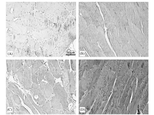
(I) Immunostaining for TNF-a: (A) negative control, (B) muscle from subject 465 yr (positive control), (C) muscle from weight maintainer, and (D) muscle from weight gainer.Anti-human TNF-a antibody (p/n 209-401-306) at 1:50. The sites of peroxidase binding were demonstrated with diamonobenzidine (p/n DAB-50). Fig 2. PMID: 16687193.
Immunohistochemistry using Rocklands polyclonal TNFa antibody showing staining of formalin/PFA-fixed paraffin-embedded sections of human artery tissue sections. Sections were fixed in formaldehyde and subjected to heat mediated antigen retrieval in citrate buffer (pH 6.0). Slides were blocked for ten minutes with 1.5% serum. Primary antibody was diluted 1:100 and incubated with samples for 24 hours at 4C. HRP-conjugated goat anti-rabbit antibody was used as the secondary antibody.
* VAT and and shipping costs not included. Errors and price changes excepted