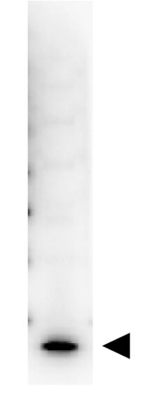
This purified antibody has been tested in western blotting and suitable for ELISA. By western blot a band approximately 17.4 kDa in size corresponding to human IL-7 protein is expected in the appropriate cell lysate or extract. Specific conditions for re
Western Blot showing detection of Human IL-7. 50ng of Human IL-7 (Lane 1) was run on a 4-20% gel and transferred to 0.45 µm nitrocellulose. After blocking with 5% Blotto (p/n B501-0500) 30 min at 20C, Anti-Human IL-7 (RABBIT) Antibody Biotin Conjugated (p/n 209-406-B94) secondary antibody was used at 1:1000 in Blocking Buffer for Fluorescent Western Blotting (p/n MB-070). HRP Streptavidin (p/n S000-03) was used at 1:40,000 in MB-070 for 30 min at 20C and imaged using the Bio-Rad VersaDoc 4000 MP. Arrow indicates correct 17 kDa molecular weight position expected for Human IL-7.
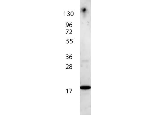
Rocklands anti-Human IL-7 antibody shows detection of a band ~17 kDa in size corresponding to recombinant human IL-7. The identity of the faint higher molecular weight band may represent a homodimer. Molecular weight markers are also shown (left). After transfer, the membrane was blocked overnight with 3% BSA in TBS followed by reaction with primary antibody at a 1:1,000 dilution. Detection occurred using peroxidase conjugated anti-Rabbit IgG (p/n 611-103-122) secondary antibody diluted 1:40,000 in blocking buffer (p/n MB-070) for 30 min at RT followed by reaction with FemtoMax(TM) chemiluminescent substrate. Image was captured using VersaDoc(TM) MP 4000 imaging system (Bio-Rad).
* VAT and and shipping costs not included. Errors and price changes excepted