Anti-EBI-3 (MOUSE) Monoclonal Antibody was produced in mouse by repeated immunizations with mature full length recombinant mouse EBI-3 produced in E.coli followed by hybridoma development.
0.02 M Potassium Phosphate, 0.15 M Sodium Chloride, pH 7.2
Form:
Lyophilized
Target:
Mouse
Antibody Type:
Primary Antibody
Application Dilute:
ELISA: 1:10,000, IP: 1µg/mL, WB: 1:1000
Application Notes:
This purified antibody has been tested for use in ELISA, IP, and Western Blot. Specific conditions for reactivity should be optimized by the end user.
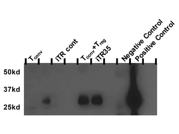
Western blot of Anti-Mouse EBI-3 Antibody. Supernatants from cultures containing indicated cells were immunoprecipitated with an anti-p35 monoclonal plus protein G beads, resolved by SDS page gel and blotted with biotinylated anti mouse-EBI-3 clone V1.4H6.29 at 1 µg/mL. The controls were supernatants from 293T cells transfected with either vector only (negative) or vector containing murine IL-35 (positive).
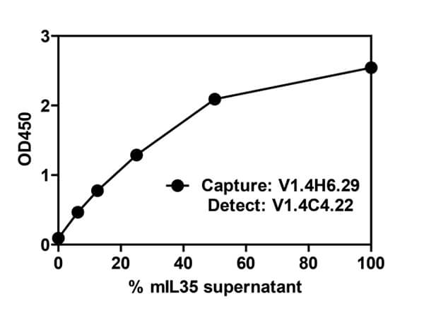
ELISA assay of EBI-3 Antibody. Supernatant from 293T cells transfected with a vector containing murine IL-35 was added to an ELISA plate coated with V1.4H6.29 (210-301-C55) at 2ug/mL in PBS. Mouse EBI-3 was detected with biotinylated V1.4C4.22 (210-301-B66).
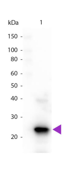
Western blot of Biotin conjugated Mouse Anti-EBI-3 (Mouse) antibody. Lane 1: Mouse EBI-3. Lane 2: None. Load: 50 ng per lane. Primary antibody: EBI-3 (Mouse) primary antibody at 1:1,000 for 60 min at RT. Secondary antibody: Peroxidase streptavidin secondary antibody at 1:40,000 for 30 min at RT. Block: MB-070 for 30 min at RT. Predicted\Observed size: 25 kDa, 25 kDa for Mouse EBI-3. Other band(s): None.
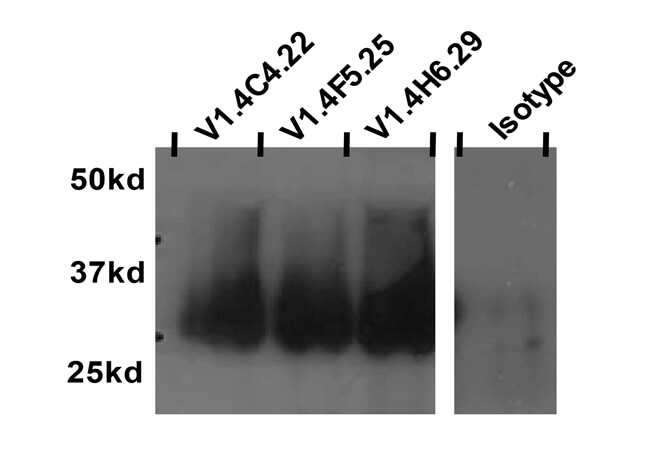
Immunoprecipitation of Mouse Anti-EBI-3 Antibody. Supernatant from 293T cells transfected with a vector containing murine IL-35 was immunoprecipitated with indicated anti-EBI-3 monoclonal clones V1.4H6.29, V1.4C4.22, and V1.4F5.25 plus Protein G beads, resolved by SDS page gel, and blotted with biotinylated anti-mouse EBI-3 clone V1.4H6.29 at 1µg/mL.
* VAT and and shipping costs not included. Errors and price changes excepted