Anti-S Opsin antibody was prepared from whole goat serum produced by repeated immunizations with a synthetic peptide corresponding to a N-terminal portion of mouse S Opsin conjugated to Keyhole Limpet Hemocyanin (KLH).
Conjugation:
Unconjugated
Alternative Names:
Goat Anti-Short-wave-sensitive opsin 1 Antibody, S opsin, Blue cone photoreceptor pigment, Blue-sensitive opsin, BOP, Short wavelength-sensitive cone opsin, Bcp, Opn1sw
0.02 M Potassium Phosphate, 0.15 M Sodium Chloride, pH 7.2
Form:
Liquid (sterile filtered)
Target:
Mouse
Antibody Type:
Primary Antibody
Application Dilute:
ELISA: 1:10,000-1:50,000, IHC: 1:100
Application Notes:
Anti-S Ospin Antibody has been tested in ELISA and IHC. Expect a band at ~39kDa in western blot using appropriate tissues and lysates. Positive control used: Mouse eye tissue in Immunohistochemistry.
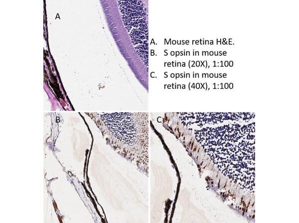
Immunohistochemistry of Goat Anti-S Opsin Antibody. Tissue: Mouse Eye Tissue (retina). Antigen Retrieval: HIER using Citrate buffer for 20mins. Fixative: none. Primary Antibody: Anti-S Ospin at 1:100 for 30 mins at RT. Secondary Antibody: Donkey Anti-Goat HRP at 4µL/mL for 20mins at RT. Counterstain: Hematoxylin. Analysis Results: S opsin is expected to stain the outer rod cells of the retina, which it does at a dilution of 1:100.
* VAT and and shipping costs not included. Errors and price changes excepted