0.02 M Potassium Phosphate, 0.15 M Sodium Chloride, pH 7.2
Form:
Lyophilized
Antibody Type:
Primary Antibody
Application Dilute:
FLISA: >1:20,000, Flow Cytometry: 1:5000, IF Microscopy: >1:5,000, WB: >1:10,000
Application Notes:
Anti-Biotin Antibody DyLight488 has been tested by Dot blot, Flow cytometry, and Immunofluorescence. The emission spectra for this DyLight(TM) conjugate match the principle output wavelengths of most common fluorescence instrumentation. This product is desi
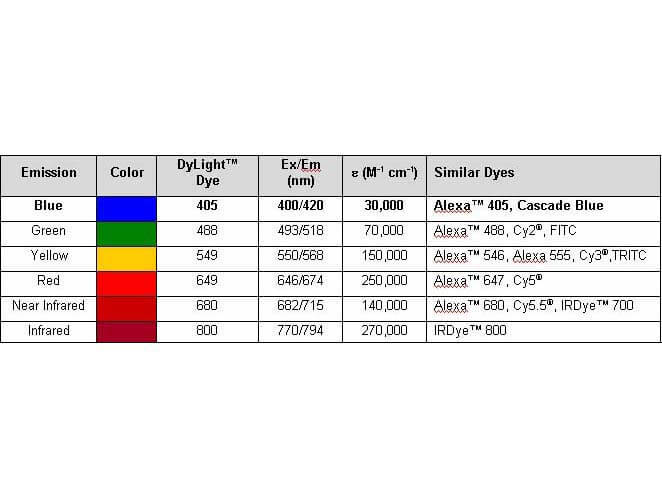
Properties of DyLight(TM) Fluorescent Dyes.
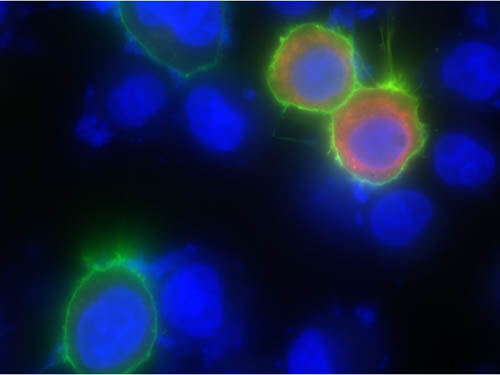
Immunofluorescence of Goat anti-biotin DyLight 488 antibody. Cells grown on glass coverslips were transiently transfected with a peroxidase-encoding plasmid and were incubated for 24 hours before carrying out a modified TSA reaction. Cells were fixed and permeabilized before probing with Rockland goat anti-biotin DyLight 488 at 1:1250 dilution for 2 hours at room temperature. Cells were washed before mounting on glass slides and imaged for biotin (green), DAPI (blue), and transfected peroxidase (red). Image courtesy Ben Dyer, University of Pennsylvania.
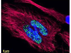
DyLight(TM) dyes can be used for multi-color immunofluorescence microscopy with uniform fluorescence intensity throughout the image. DyLight(TM) dyes are exceptionally bright and photostable and are optimized for microscopy and microarray detection methods. This image shows anti-histone detection using a DyLight(TM) 488 conjugate (green). Anti-Tubulin was detected using a DyLight(TM) 549 conjugate (red). Nuclei were counter-stained using DAPI (blue). The image was captured using an Axio Imager.Z1 (Zeiss Micro Imaging Inc).
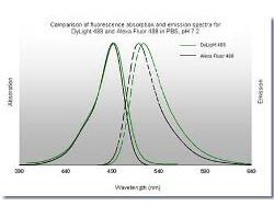
DyLight 488 Fluorescence Spectra.
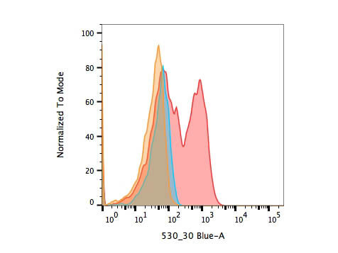
Flow Cytometry of Goat anti-biotin DyLight 488 antibody. Cells were transiently transfected with a peroxidase-encoding plasmid and were incubated for 24 hours before carrying out a modified TSA reaction. Cells were fixed and permeabilized before probing with Rockland goat anti-biotin DyLight 488 at 1:5000 dilution for 2 hours at room temperature. Transfected and biotin supplemented population (red) is compared to transfected, non-supplemented (blue) and non-transfected, biotin supplemented (orange) controls. Image courtesy Ben Dyer, University of Pennsylvania.
* VAT and and shipping costs not included. Errors and price changes excepted