0.02 M Potassium Phosphate, 0.15 M Sodium Chloride, pH 7.2
Form:
Lyophilized
Antibody Type:
Primary Antibody
Application Dilute:
FLISA: >1:20,000, IF Microscopy: >1:5,000, WB: 1:10,000 - 1:25,000
Application Notes:
This product is designed for immunofluorescence microscopy, fluorescence based plate assays (FLISA) and fluorescent western blotting. This product is also suitable for multiplex analysis, including multicolor imaging, utilizing various commercial platfor
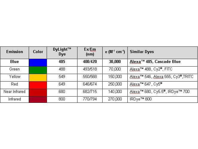
Properties of DyLight(TM) Conjugates.
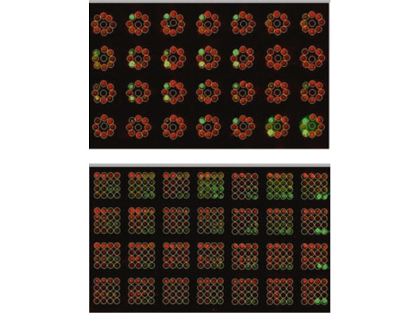
Quantification of SH2 binding with ImageStudio using Anti-GST DyLight(TM)800. (Top) Analysis is set to 3*3 arrays and spots are rearranged for alignment with the circular MBA plate array. (Bottom) Analysis is set to 4*4 arrays and spots are aligned with the square MWA plate array. Both SH2 binding (800 nm channel) and peptide loading (700 nm channel) are quantified at the same time. Each set of samples is incubated with different GST-SH2 probes in separate wells of a 96-well chamber plate. SH2 binding is detected via 800 nm infrared dye-conjugated anti-GST antibody and IR imager. The loaded amount of biotin-tagged peptides can be monitored using 680 nm infrared dye-conjugated avidin. PMID: 28092049.
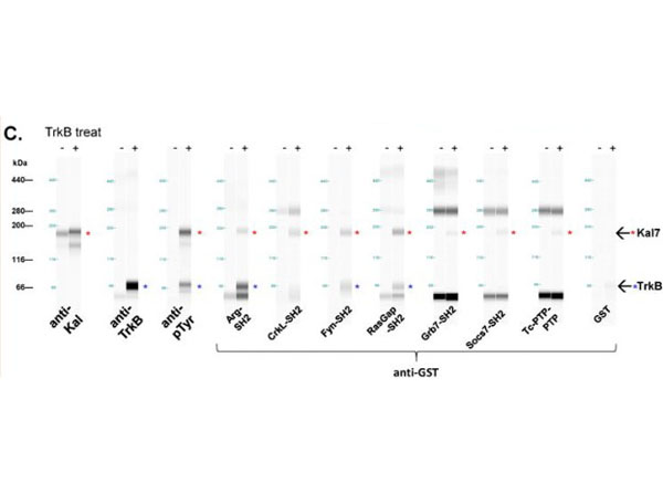
Far-Western analysis of selected interactors using Anti-GST DyLight(TM)800. C.Validation of SH2 screen. Capillary far-Western assay was performed to confirm direct interactions between the probe and purified, phosphorylated bKal7. Red asterisks in blot view indicate the presence of phospho-Kal7 (190 KDa) with TrkB treatment, blue asterisks indicate TrkB (70 KDa). The higher molecular weight bands detected by several probes are non-specific.Fig 6. PMID: 28418645.
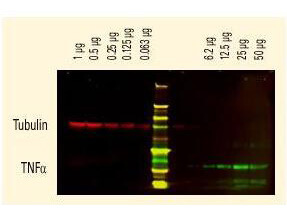
DyLight(TM) dyes can be used for two-color Western Blot detection with low background and high signal. Anti-tubulin was detected using a DyLight(TM) 680 conjugate. Anti-TNFa was detected using a DyLight(TM) 800 conjugate. The image was captured using the Odyssey Infrared Imaging System developed by LI-COR.
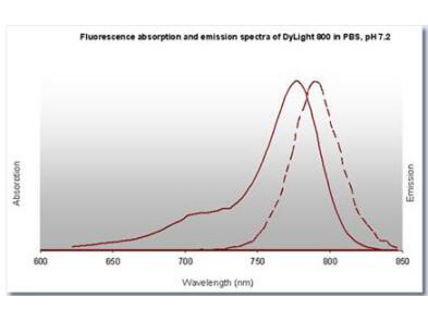
DyLight(TM) 800 Fluorescence Spectra.
* VAT and and shipping costs not included. Errors and price changes excepted