Recombinant Green Fluorescent Protein (GFP) fusion protein corresponding to the full length amino acid sequence (246 aa) derived from the jellyfish Aequorea victoria.
Conjugation:
Unconjugated
Alternative Names:
mouse anti-GFP antibody, Green Fluorescent Protein, GFP antibody, Green Fluorescent Protein antibody, EGFP, enhanced Green Fluorescent Protein, Aequorea victoria, Jellyfish
0.02 M Potassium Phosphate, 0.15 M Sodium Chloride, pH 7.2
Form:
Liquid (sterile filtered)
Antibody Type:
Primary Antibody
Application Dilute:
ELISA: 1:10,000 - 1:30,000, Flow Cytometry: User Optimized, IHC: 1:1,000 - 1:5,000, IF Microscopy: User Optimized, WB: 1:3,000 - 1:30,000
Application Notes:
Monoclonal anti-GFP is designed to detect enhanced GFP and GFP containing recombinant proteins. Tested in ELISA, IP, and WB and suitable in FACS, IHC, IF. This antibody can be used to detect GFP by ELISA (sandwich or capture) for the direct binding of an
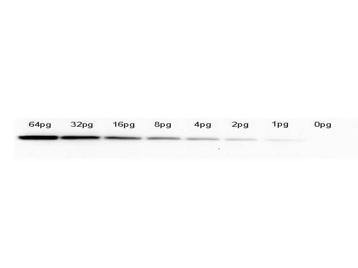
Western Blot of anti-GFP monoclonal antibody. Lane 1: 64pg of recombinant GFP protein (p/n 000-001-215) were spiked into a HeLa cell-derived lysates (p/n W09-000-364). Lane 2: 32pg of recombinant GFP protein were spiked into a HeLa cell-derived lysates. Lane 3: 16pg of recombinant GFP protein were spiked into a HeLa cell-derived lysates. Lane 4: 8pg of recombinant GFP protein were spiked into a HeLa cell-derived lysates. Lane 5: 4pg of recombinant GFP protein were spiked into a HeLa cell-derived lysates. Lane 6: 2pg of recombinant GFP protein were spiked into a HeLa cell-derived lysates. Lane 7: 1g of recombinant GFP protein were spiked into a HeLa cell-derived lysates. Lane 8: 0pg of recombinant GFP protein were spiked into a HeLa cell-derived lysates. Primary antibody: anti-GFP monoclonal antibody at 1:400 for overnight at 4C. Secondary antibody: HRP-conjugated anti-Mouse IgG (p/n 610-4302) was performed at a dilution of 1:20,000 for 1h at 4C. Block: TTBS (p/n MB-013) supplemented with 1% BSA (p/n BSA-50) for 1 h at 4C. Predicted/Observed size: 27 kDa for GFP. Other band(s): none.
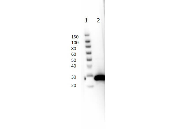
Western blot of Mouse Anti-GFP Antibody. Lane 1: Thermo SuperSignal Molecular Weight Marker. Lane 2: GFP protein (p/n 000-001-215) [50ng]. Primary Antibody: Anti-GFP at 1:1000 overnight at 2-8C. Secondary Antibody: Rabbit Anti-Mouse IgG HRP (p/n 610-4302) at 1:40,000 for 30mins at RT. Block: BlockOut Buffer (p/n MB-073). Expected MW: ~27kDa.
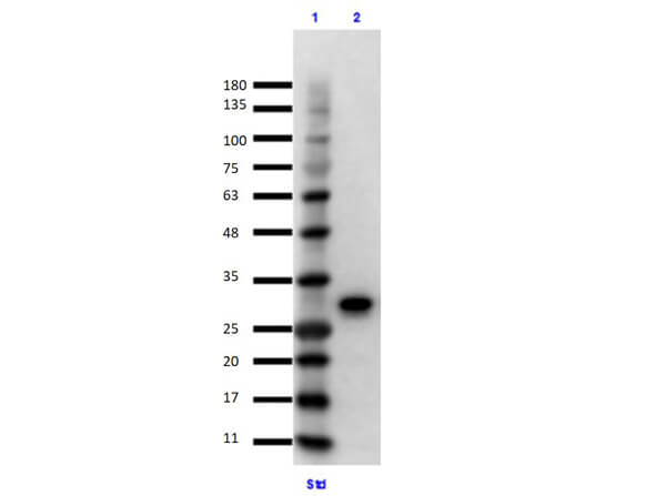
Immunoprecipitation/Western Blot using GFP Protein. Lane 1: Opal Prestained Molecular Weight Marker (p/n MB-210-0500). Lane 2: GFP Input (p/n 000-001-215) Reduced [10µL]. Primary IP Antibody: Mouse Anti-GFP (p/n 600-301-215) at 10µg overnight at 2-8C. Secondary Antibody: TrueBlot Anti-Mouse Ig IP Agarose Beads (p/n 00-8811-25) at 500µg for 1hr at RT. Buffer: BlockOut Buffer (p/n MB-073) for 30 mins at RT. Exposure: 7 sec.
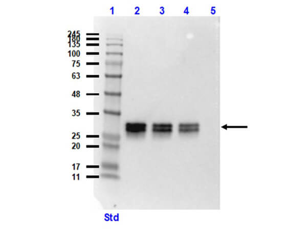
Western blot of Mouse Anti-GFP Antibody. Lane 1: Opal Prestained Molecular Weight Marker (p/n MB-210-0500). Lane 2: HeLa WC Lysate+GFP protein (p/n W09-000-364 [10µg]/ p/n 000-001-215 [50ng]). Lane 3: HeLa WC Lysate+GFP protein (10µg/20ng). Lane 4: HeLa WC Lysate+GFP protein (10µg/10ng). Lane 5: HeLa Whole Cell Lysate (p/n W09-000-364) (10µg). Primary Antibody: Anti-GFP at 1:1000 overnight at 2-8C. Secondary Antibody: Rabbit Anti-Mouse IgG HRP (p/n 610-4302) at 1:40,000 for 30mins at RT. Block: BlockOut Buffer (p/n MB-073). Expected MW: ~27kDa.
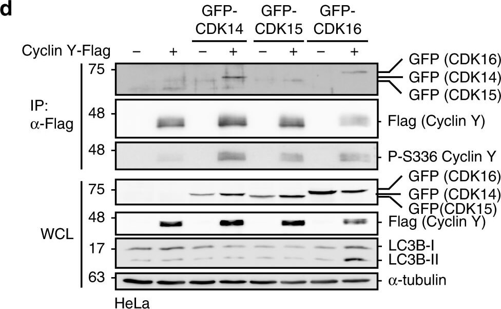
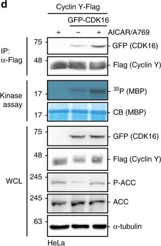
Protein microarray screen for the identification of AMPK substrates.a Schematic representation of the ProtoArray based screen with approximately 9000 human proteins using AMPK (see also Supplementary Data 1). b Details of two sub-arrays incubated with or without AMPK with marked substrates are shown. c GST-CDK16, Cyclin Y-His6 and GST were incubated in the presence of [gamma-32P]-ATP with AMPK. Phosphorylation was determined by autoradiography (32P, top). Proteins were visualized by Coomassie blue staining (CB, bottom, n=2). d HeLa cells were transfected with vectors expressing GFP-CDK16 and Cyclin Y-Flag and treated for 1h with 0.5mM AICAR/50µM A769662 (A769) as indicated. Cyclin Y-Flag was immunoprecipitated with Flag antibodies (IP) and immunoblotted against CDK16 and Cyclin Y or used for in vitro kinase assays with myeloid basic protein (MBP) as substrate. Autoradiographs (32P) and Coomassie blue staining (CB) of MBP are displayed. Whole cell lysates (WCL) were immunoblotted with the indicated antibodies (n=3). e Quantification of CDK16 co-immunoprecipitated with Cyclin Y. Statistical significance was measured via unpaired and two-tailed Students t-tests and is presented as follows: **p<0.01, and ***p<0.001. All error bars indicate SD (n=3, Cyclin Y+AICAR/A769 vs. Cyclin Y/CDK16: t=8.719, df=4, Cyclin Y/CDK16 vs. Cyclin Y/CDK16+AICAR/A769: t=5.595, df=4). n biological independent replicate. SD standard deviation. Source data are provided as a Source Data file. Figure provided b
* VAT and and shipping costs not included. Errors and price changes excepted