Anti-Green Fluorescent Protein (GFP) is produced by immunizing GFP containing fusion protein that corresponds to the full length amino acid sequence (246aa) derived from the jellyfish Aequorea victoria.
Conjugation:
Biotin
Alternative Names:
mouse anti-GFP antibody biotin conjugation, biotin conjugated mouse anti-GFP antibody, Green Fluorescent Protein, GFP antibody, Green Fluorescent Protein antibody, EGFP, enhanced Green Fluorescent Protein, Aequorea victoria, Jellyfish
Monoclonal anti-GFP is designed to detect enhanced GFP and GFP containing recombinant proteins. Tested in E, WB, IHC. This antibody can be used to detect GFP by ELISA (sandwich or capture) for the direct binding of antigen. Biotin conjugated monoclonal a
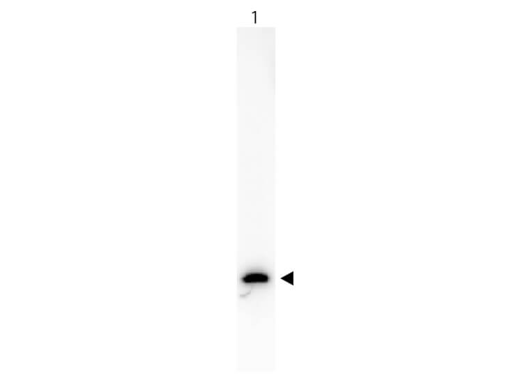
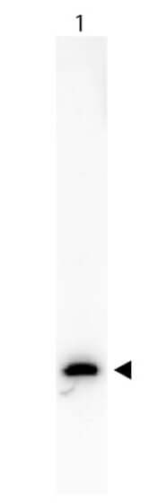
Western Blot of Anti-GFP (MOUSE) Monoclonal Antibody Biotin Conjugated antibody. Lane 1: 50ng of GFP. Lane 2: none. Primary antibody: none. Secondary antibody: Anti-GFP (MOUSE) Monoclonal Antibody Biotin Conjugated (p/n 600-306-215) secondary antibody was used at 1:5000 in Blocking Buffer for Fluorescent Western Blotting (p/n MB-070) for 45 min at RT. HRP Streptavidin (p/n S000-03) was used at 1:40,000 in MB-070 for 30 min at 20C. Block: 5% Blotto (p/n B501-0500) 30 min at 20C. Predicted/Observed size: 28 kDa for GFP. Other band(s): none.
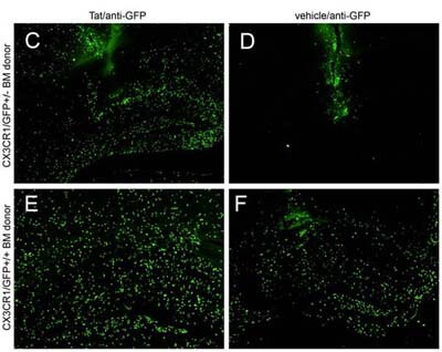
IHC - Immuno-Fluorescence of Biotin Mouse anti-GFP antibody. Biotin mouse anti GFP used 1:5000 As referenced in: Lu S-M, Tremblay M-E , King IL, Qi J, Reynolds HM, et al. (2011) HIV-1 Tat-Induced Microgliosis and Synaptic Damage via Interactions between Peripheral and Central Myeloid Cells. PLoS ONE 6(9): e23915. doi:10.1371/journal.pone.0023915
* VAT and and shipping costs not included. Errors and price changes excepted