0.02 M Potassium Phosphate, 0.15 M Sodium Chloride, pH 7.2
Form:
Liquid (sterile filtered)
Target:
Human
Antibody Type:
Primary Antibody
Application Dilute:
ELISA: 1:5,000 - 1:25,000, WB: 1:500 - 1:3,000
Application Notes:
IKKepsilon pT501 antibody is tested in ELISA, western blotting, and although not tested, this antibody is likely functional in immunohistochemistry and immunoprecipitation. An 85 kDa band corresponding to human IKKe is detected. HeLa cells or TNF inducible KBM
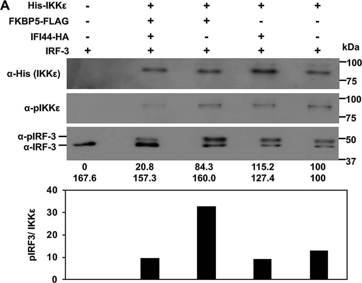
IFI44 decreases the kinase activity of IKKbeta and IKKepsilon. Human 293T cells were silenced for IFI44, or for FKBP5, and were transfected with plasmids expressing His-IKKepsilon (A) or MYC-IKKbeta (B), together with IFI44-HA, and FKBP5-FLAG expression plasmids. At 24 hpt, IKKepsilon (A) and IKKbeta (B) complexes were purified with anti-His and anti-MYC antibodies, respectively, and these complexes were assayed in kinase assays using IRF-3 (for the IKKepsilon complexes shown in panel A) and IkBalpha (for the IKKbeta complexes shown in panel B) as substrates. The levels of phosphorylated and unphosphorylated forms of IRF-3 (panel A, bottom blot) and IkBalpha (panel B, third and fourth blots) were analyzed by Western blotting using specific antibodies. Levels of IKKepsilon were analyzed using an anti-His-specific antibody (A, first blot) and anti-pIKKepsilon (A, second blot), and levels of IKKbeta were analyzed using an anti-MYC-specific antibody (B, first blot) and anti-pIKKbeta (B, second blot). Western blots were quantified by densitometry using ImageJ software (v1.46). Protein expression levels in cells expressing IKKepsilon (A) and IKKbeta (B) alone were assigned a value of 100% for comparisons with the levels of expression in cells expressing the different combinations of IKKepsilon/IFI44/FKBP5 (A) or IKKbeta/IFI44/FKBP5 (B) (numbers are indicated below each plot). pIRF-3 and IRF-3 levels (observed in the same bottom blot in panel A) and pIkBalpha and IkBalpha (third and bottom blot in panel B) are represented with numbers below each blot. Levels of pIRF-3 and pIkBalpha normalized to the levels of IKKepsilon and IKKbeta are represented in the bottom graphs in panels A and B, respectively. Molecular weight markers are indicated (in kilodaltons) on the right. Figure provided by CiteAb. Source: MBio, PMID: 31455651.
* VAT and and shipping costs not included. Errors and price changes excepted