
AKT phospho T308 Antibody, Rabbit, Polyclonal
Catalog Number:
ROC-600-401-269
- Images (9)

| Article Name: | AKT phospho T308 Antibody, Rabbit, Polyclonal |
| Biozol Catalog Number: | ROC-600-401-269 |
| Supplier Catalog Number: | 600-401-269 |
| Alternative Catalog Number: | ROC-600-401-269 |
| Manufacturer: | Rockland Immunochemicals |
| Host: | Rabbit |
| Category: | Antikörper |
| Application: | DOT, ELISA, IHC, WB |
| Species Reactivity: | Human, Mouse, Rat |
| Immunogen: | Anti-AKT pT308 polyclonal antibody was produced by repeated immunizations with a phosphorylated synthetic peptide corresponding to residues surrounding threonine 308 of human AKT1 protein. |
| Conjugation: | Unconjugated |
| Alternative Names: | rabbit anti-AKT pT308 Antibody, AKT1 phospho T308, RAC-PK-alpha, Protein kinase B, PKB, C-AKT, RAC-alpha serine/threonine-protein kinase, Proto-oncogene c-Akt, AKT1, AKT 1, AKT-1 |
| Application Dilute: | ELISA: 1:15,000, Flow Cytometry: User Optimized, IHC: 1:200, WB: 1:1000 |
| Application Notes: | AKT antibody is phospho specific for pT308 and is tested for western blotting, Immunohistochemistry, dot blot, and ELISA. This antibody is suitable for flow cytometry. |
|
|
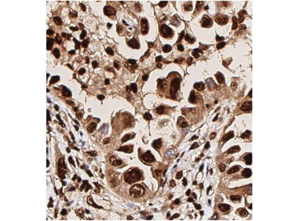
Immunohistochemistry of Rabbit Anti-AKT pT308 Antibody. Tissue: human lung tissue. Antigen retrieval: HIER using citrate buffer for 20 minutes. Fixative: None. Primary Antibody: Anti-AKT phosphoT308 at 1:200 for 30 minutes at RT. Secondary Antibody: Anti-Rabbit Poly-HRP-IgG Ready to Use for 8 minutes at RT. Counterstain: Hematoxylin. Substrate: DAB. Analysis: Strong staining in nucleus. May be suitable with more dilutions. |
|
|
|
|
|
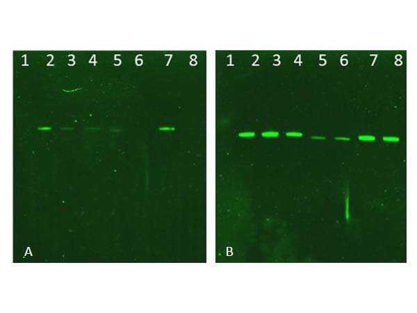
Western Blot of Rabbit AKT Antibodies. Lane 1: NIR MW protein ladder. Lane 2: AKT1, recombinant: 009-001-P21. Lane 3: AKT1, phosphatase-treated: 009-001-I51. Lane 4: AKT1, mutant T308A/S473A: 009-001-P22. Lane 5: AKT2, recombinant: 009-001-P23. Lane 6: AKT2, phosphatase-treated: 009-001-E71. Lane 7: AKT3, recombinant: 009-001-P24. Lane 8: AKT3, phosphatase-treated: 009-001-E75. Load: 50ng per lane. Blot A: 600-401-269 Anti-Akt pT308 used at 1:2270, Blot B: 100-401-401 Anti-Akt used 1:1000. |
|
|
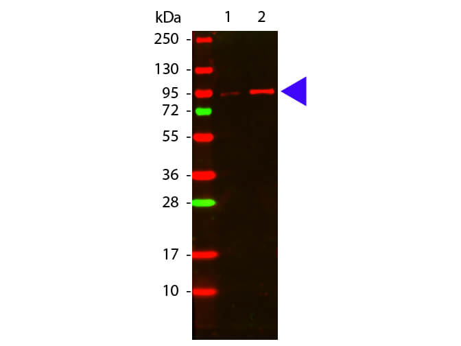
Western Blot of Rabbit anti-Akt phospho T308 antibody. Lane 1: GST tagged AKT1 un-active recombinant protein. Lane 2: GST tagged AKT1 active recombinant protein. Load: 50 ng per lane. Primary antibody: Akt phospho T308 antibody at 1:1,000 for overnight at 4C. Secondary antibody: DyLight(TM) 649 rabbit secondary antibody at 1:20,000 for 30 min at RT. Block: MB-070 for 30 min at RT. Other band(s): none. |
|
|
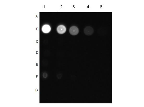
Dot Blot of Rabbit Anti-AKT pT308 Antibody. Dilutions in Columns: (1) 100ng, (2) 33.33ng, (3) 11.11ng, (4) 3.7ng, (5) 1.23ng. Tested BSA Peptide Reactivity in Rows: (A) AKT1-BSA, (B) AKT1 pT308-BSA, (C) AKT1 S473-BSA, (D) AKT1 pS473-BSA, (E) CDC27 T244-BSA, (F) CDC27 pT244-BSA, (G) BSA control. Primary Antibody: Anti-AKT pT308 at 1µg/mL overnight at 2-8C. Secondary Antibody: Goat anti-Rabbit IgG HRP (p/n 611-103-122) at 1:70,000 at RT for 30mins. Block: BlockOut Buffer (p/n MB-073). |
|
|
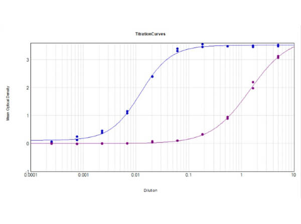
ELISA Results of Rabbit Anti-AKT pT308 Antibody tested against BSA-conjugated non-phospho [purple] and phospho [blue] forms of immunizing peptide. Each well was coated in duplicate with either 0.1µg of conjugate. The working dilution is 1:81,300. The starting dilution of antibody was 5µg/ml and the X-axis represents the Log10 of a 3-fold dilution. This titration is a 4-parameter curve fit where the IC50 is defined as the tite |
|
|
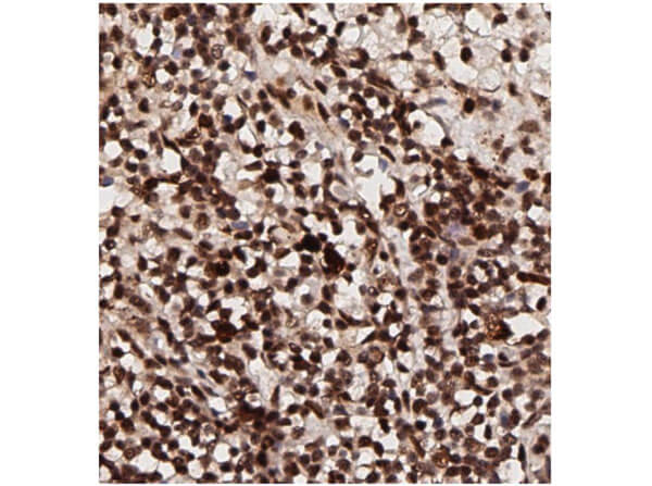
Immunohistochemistry of Rabbit Anti-AKT pT308 Antibody. Tissue: human breast tissue (lymph nodes). Antigen retrieval: HIER using citrate buffer for 20 minutes. Fixative: None. Primary Antibody: Anti-AKT phosphoT308 at 1:200 for 30 minutes at RT. Secondary Antibody: Anti-Rabbit Poly-HRP-IgG Ready to Use for 8 minutes at RT. Counterstain: Hematoxylin. Substrate: DAB. Analysis: Strong staining in nucleus. May be suitable with more dilutions. |
|
|
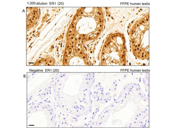
Immunohistochemistry of Rabbit Anti-AKT pT308 Antibody. Tissue: human testis tissue. Antigen retrieval: Heat induced antigen retrieval was performed using Leica Bond Epitope Retrieval Buffer 1 (Citrate solution, pH6.0) for 20 minutes. Fixative: None. Primary Antibody: (A). Anti-AKTpT308 at 1:200 for 30 minutes at RT. (B). Negative control. Secondary Antibody: Anti-Rabbit Poly-HRP-IgG Ready to Use for 8 minutes at RT. Counterstain: Hematoxylin. Substrate: DAB. Analysis: Cells in seminiferous ducts and Leydig cells show moderate cytoplasmic staining. |
|
|
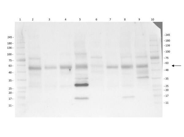
Multi-Lysate Western Blot of Rabbit Anti-AKT pT308 Antibody. Lane 1: Opal Pre-stained Molecular Weight Marker (p/n MB-210-0500). Lane 2: Human Spleen Lysate. Lane 3: Hu Small Intestine Lysate. Lane 4: Hu Placenta Lysate. Lane 5: Hu Skeletal Muscle Lysate. Lane 6: Hu Brain Cerebellum Lysate. Lane 7: Hu Lung Lysate. Lane 8: Hu Tonsil Lysate. Lane 9: Hu Thymus Lysate. Lane 10: Opal Pre-stained Molecular Weight Marker (p/n Mb-210-0500). Primary Antibody: Anti-AKT pT308 at 1:1000 overnight at 2-8C. Secondary Antibody: Goat Anti-Rabbit IgG HRP (p/n 611-103-122) at 1:40,000 at RT for 60mins. Block: BlockOut Buffer (p/n MB-073). Predicted MW: ~55kDa. Observed MW: ~28, ~58kDa. Notes: Ubiquitous. |
Product Guarantee and Expert Support
