
RFP Antibody Pre-adsorbed, Rabbit, Polyclonal
Catalog Number:
ROC-600-401-379
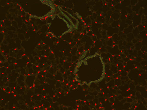
- Images (46)


| Article Name: | RFP Antibody Pre-adsorbed, Rabbit, Polyclonal |
| Biozol Catalog Number: | ROC-600-401-379 |
| Supplier Catalog Number: | 600-401-379 |
| Alternative Catalog Number: | ROC-600-401-379 |
| Manufacturer: | Rockland Immunochemicals |
| Host: | Rabbit |
| Category: | Antikörper |
| Application: | ELISA, IF, IHC, WB |
| Species Reactivity: | Other |
| Immunogen: | The immunogen is a Red Fluorescent Protein (RFP) fusion protein corresponding to the full-length amino acid sequence (234aa) derived from the mushroom anemone Discosoma. |
| Conjugation: | Unconjugated |
| Alternative Names: | rabbit anti-RFP antibody, DsRed, rDsRed, Discosoma sp. Red Fluorescent Protein, Red fluorescent protein drFP583. |
| Application Dilute: | ELISA: 1:28,700 - 1:48,700, Flow Cytometry: 1:200 - 1:2,000, IHC: 1:200 - 1:2,000, IF Microscopy: 1:200 - 1:2,000, IP: User Optimized, WB: 1:1,000 - 1:5,000 |
| Application Notes: | Polyclonal anti-RFP is designed to detect RFP and its variants. This antibody has been tested by ELISA, Western blot, IF, and IHC, and is suitable for use in EM, FC, FISH, IP, and multiplex assays based on published references. This antibody can be used |
|
|
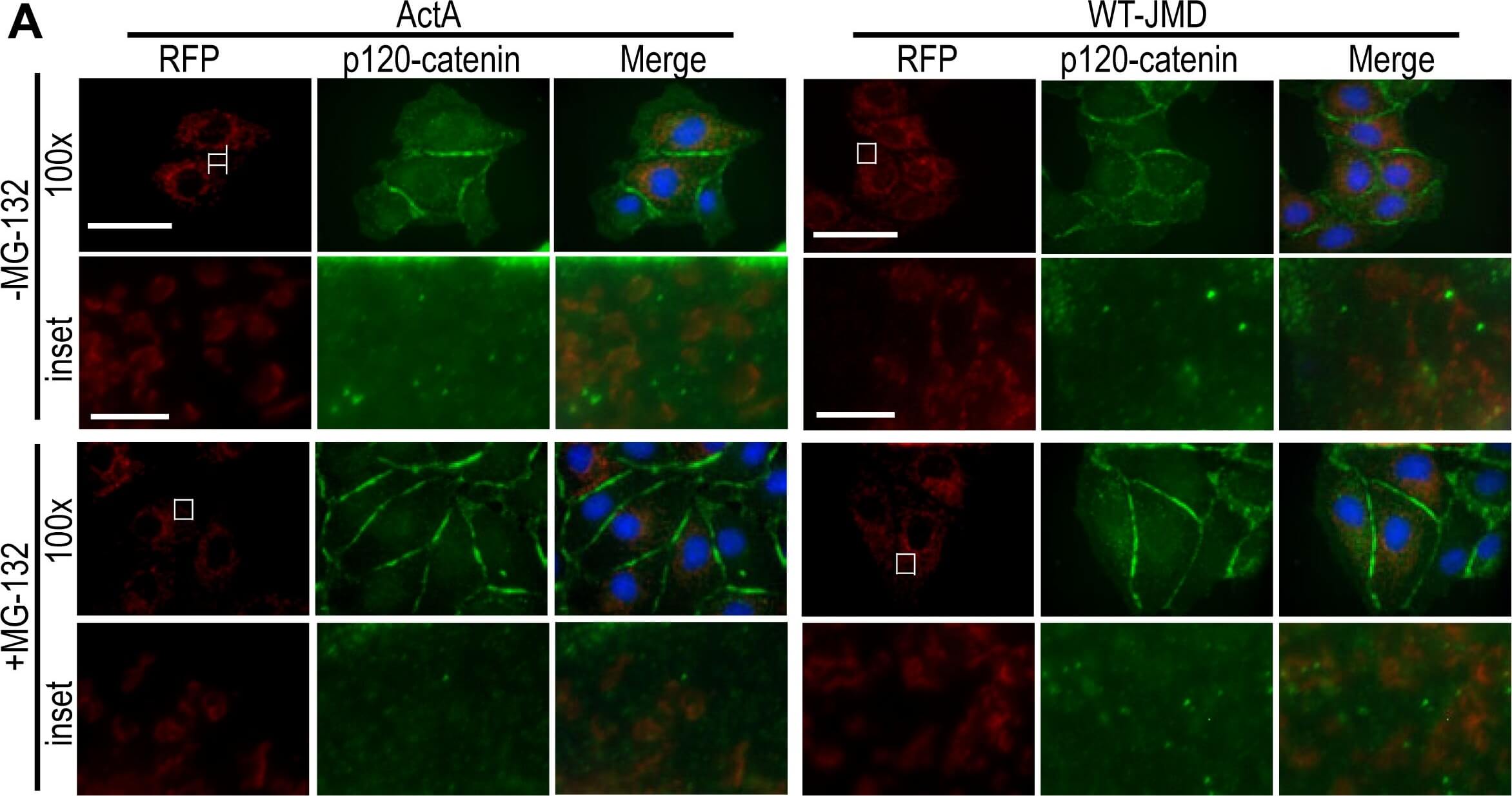
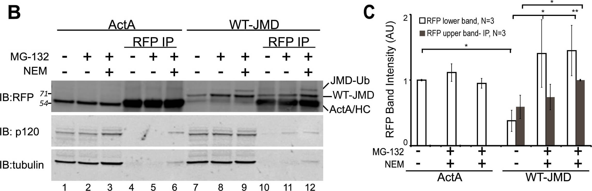
Localization and binding of WT-JMD and p120-catenin.(A) Immunofluorescence of MDCK cells transiently expressing ActA or WT-JMD. Images for RFP (red), p120-catenin (green) and merged are shown separately (100*). Boxed areas are shown as higher magnifications below (RFP-, p120- and merge-inset). All images were from the same experiment and processed identically between cell lines. Scale bar is 25 µm in 100* images, and 5 µm in insets. (B) Lysates and RFP immunoprecipitates (IP) of ActA and WT-JMD stable cell lines under normal conditions, upon proteasome inhibition, or NEM treatment (to inhibit de-ubiquitinating enzymes). Immunoblots (IB) for RFP show: a... |
|
|
|
|
|
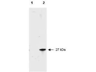
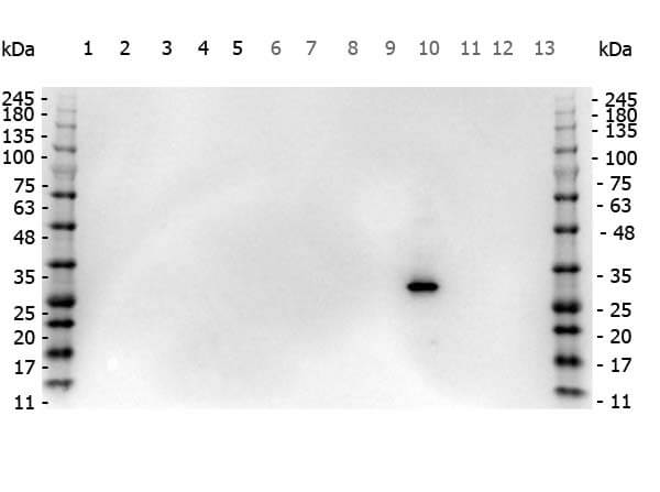
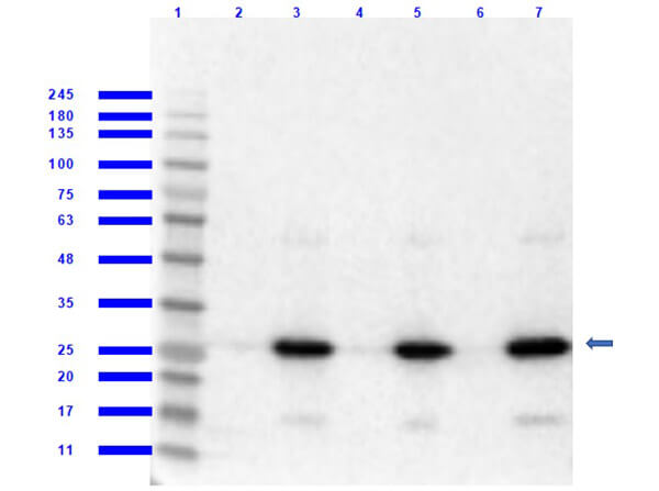
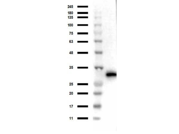
Western blot of RFP recombinant protein detected with Rocklands polyclonal anti-RFP antibody. Lane 1 shows no reaction against a GFP recombinant protein present in 10 µg of HeLa cell extract. Lane 2 shows a single band detected in 10 µg of a HeLa lysate containing RFP recombinant protein as a 27 kDa band. A 4-12% Bis-Tris gradient gel (Invitrogen) was used for SDS-PAGE. The membrane was blocked and then probed with Anti-RFP diluted 1:2,500 for 1 h at RT followed by washes and reaction with a 1:5,000 dilution of IRDye(TM)800 conjugated Goat-a-Rabbit IgG [H&L] MX (611-132-122). IRDye(TM)800 fluorescence image was captured using the Odyssey Infrared Imaging... |
|
|
|
|
|
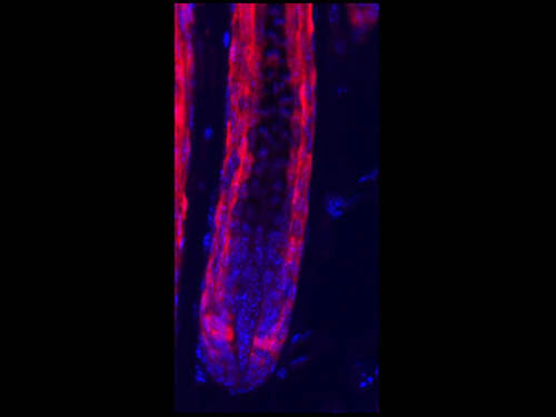
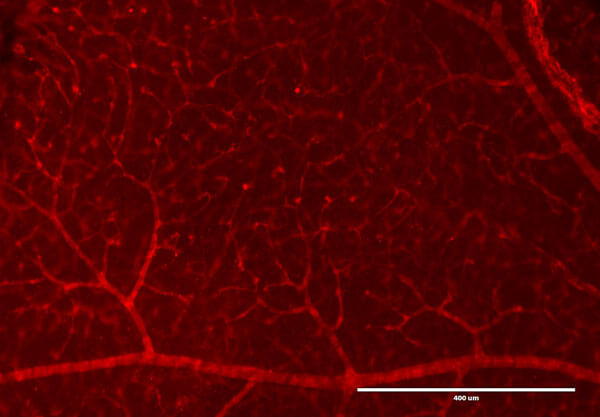
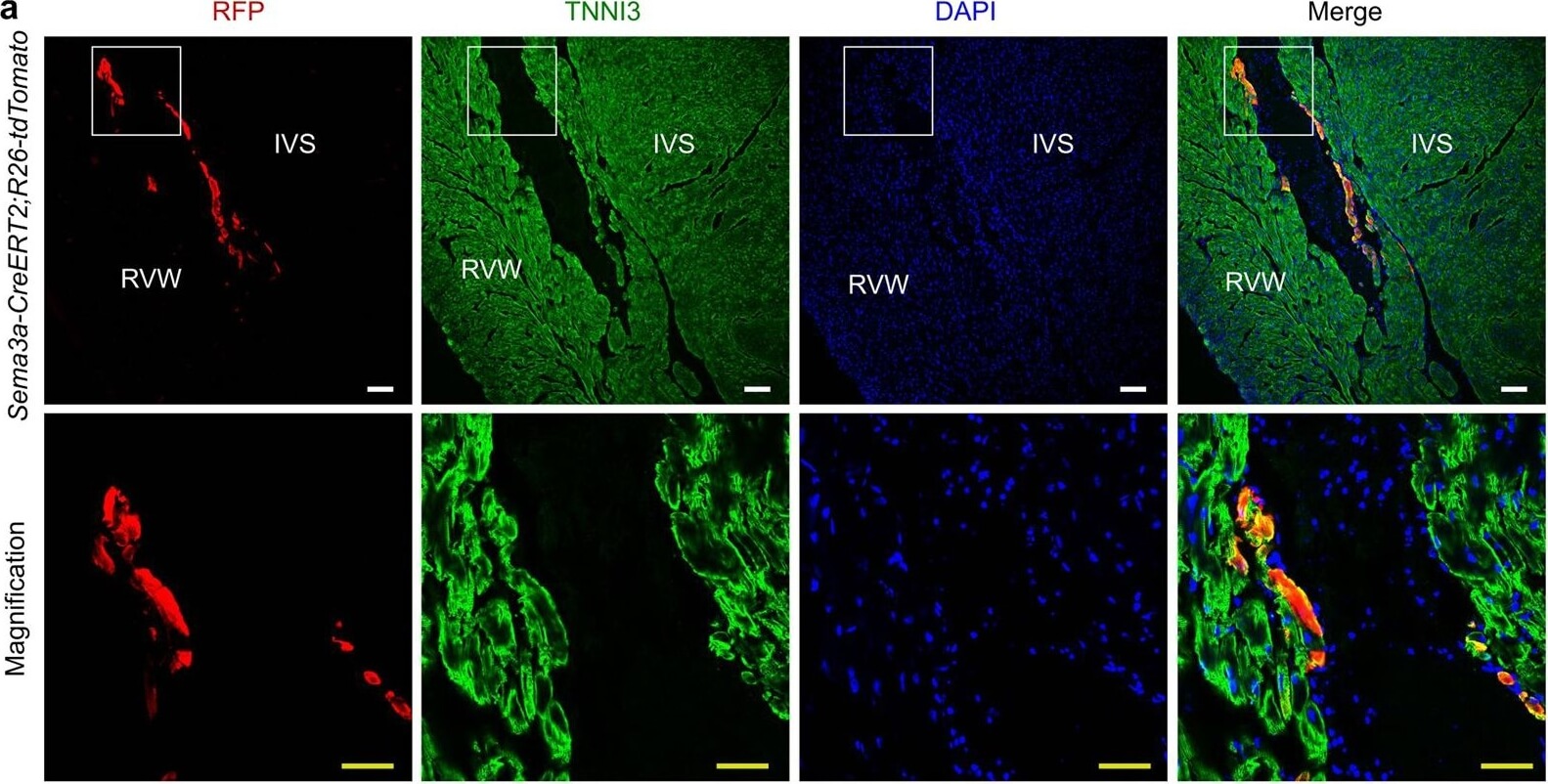
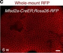
Immunofluorescence Microscopy of Rabbit Anti-RFP antibody. Tissue: HopERCre/+, R26Tom/+ mice. Fixation: 0.5% PFA. Antigen retrieval: Tamoxifen. Primary antibody: RFP antibody at 10 µg/mL for 1 h at RT. Secondary antibody: Fluorescein rabbit secondary antibody at 1:10,000 for 45 min at RT. Localization: RFP is nuclear and occasionally cytoplasmic. Staining: Hop-derived cells in the hair follicle, labeled in red. Courtesy of Rajan Jain at UPenn. |
|
|
|
|
|
|
|
|
|
|
|
|
|
|
|
|
|
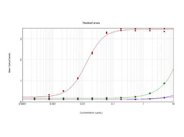
ELISA Results of Polyclonal Rabbit Anti-RFP Antibody tested against purified RFP protein. Each well was coated in duplicate with 1.0 µg of RFP (p/n 000-001-379) [red], Human IgG (p/n 009-0102) [green], Mouse IgG (p/n 010-0102) [blue], Rat IgG (p/n 012-0102) [purple]. The starting dilution of antibody was 5µg/mL and the X-axis represents the Log10 of a 3-fold dilution. This titration is a 4-parameter curve fit where the IC50 is defined as the titer of the antibody. Assay performed using 1% Fish Gel in PBS Blocking Buffer (p/n MB-066), Goat Anti-Rabbit IgG HRP conjugated (p/n 611-103-122) and TMB substrate (p/n TMBE-1000). Results show RFP titer >75,000,... |
|
|
|
|
|
|
|
|
|
|
|
|
|
|
|
|
|
|
|
|
|
|
|
|
|
|
|
|
|
Preserved cell-type specific expression patterns in BAC transgenic mice created with advanced recombineering strategies. (A-D) Anti-2A slice staining in brain slices from DAT-ChETA line 3 mice reveals the distribution of membrane-targ |
|
|
|
|
|
|
|
|
|
|
|
|
|
|
|
|
|
|
|
|
|
|
|
|
|
|
|
|
|
|
|
|
|
|
|
|
|
|
|
|
|
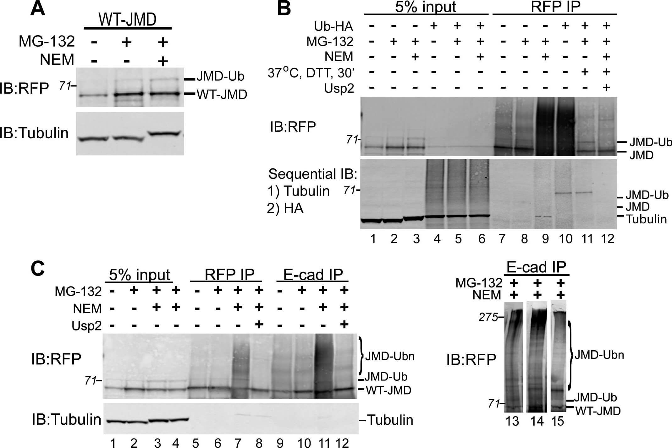
E-cadherin JMD is Ubiquitinated.(A) Lysates of WT-JMD stable cell lines under normal conditions, upon proteasome inhibition (MG-132), or inhibition of deubiquitinating enzymes (NEM). Immunoblot (IB) for RFP shows a slower migrating band (JMD-Ub) in the presence of MG-132 and NEM. (B) In a separate experiment MDCK cells stably expressing WT-JMD were transiently transfected with Ub-HA where indicated. Cells were extracted and RFP immunoprecipitations were preformed under normal conditions, upon proteasome inhibition, NEM treatment (to inhibit de-ubiquitinating enzymes), addition of the deubiquitinating enzyme Usp2, or mock transfections. DTT was added to... |
|
|
|
|
|
|
|
|
|
|
|
|
|
|
|
|
|
|
|
|
|
|
|
|
|
|
|
|
|
Product Guarantee and Expert Support
