The immunogen is a Red Fluorescent Protein (RFP) fusion protein corresponding to the full-length amino acid sequence (234aa) derived from the mushroom anemone Discosoma.
Conjugation:
Unconjugated
Alternative Names:
DsRed protein, rDsRed, Discosoma sp. Red Fluorescent Protein, Red fluorescent protein drFP583, sea anemone Discosoma sp. Mushroom, RFP antibody, RFP-RTU, Ready To Use Antibody, RTU Antibody
0.02 M Potassium Phosphate, 0.15 M Sodium Chloride, pH 7.2
Form:
Liquid (sterile filtered)
Antibody Type:
Primary Antibody
Application Dilute:
ELISA: User Optimized, IHC: User Optimized, IF Microscopy: User Optimized, IP: User Optimized, WB: 1:1,000
Application Notes:
Ready-To-Use Anti-RFP is designed to detect RFP and its variants. Ready-To-Use Anti-RFP Rabbit Polyclonal Antibody has been optimized and tested in ELISA and in Western blot using 1:1000 dilution. This Anti-RFP (RTU) Antibody is sufficient to run 10 West
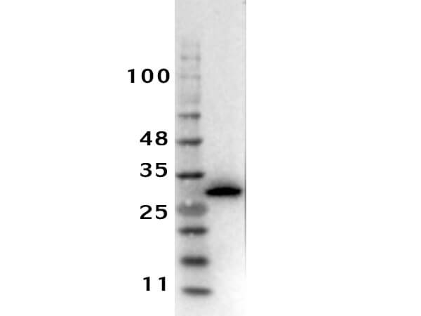
Western Blot Results of Ready to Use Rabbit Anti-RFP Antibody. Lane 1: recombinant RFP. Load: 0.05µg. Primary Antibody: Ready to Use Rabbit Anti-RFP Antibody 1:1000 overnight at 4C. Secondary Antibody: Donkey Anti-Goat HRP (p/n 605-703-125) at 1:40,000 for 30 min RT. Block: BlockOut Buffer (p/n MB-073).
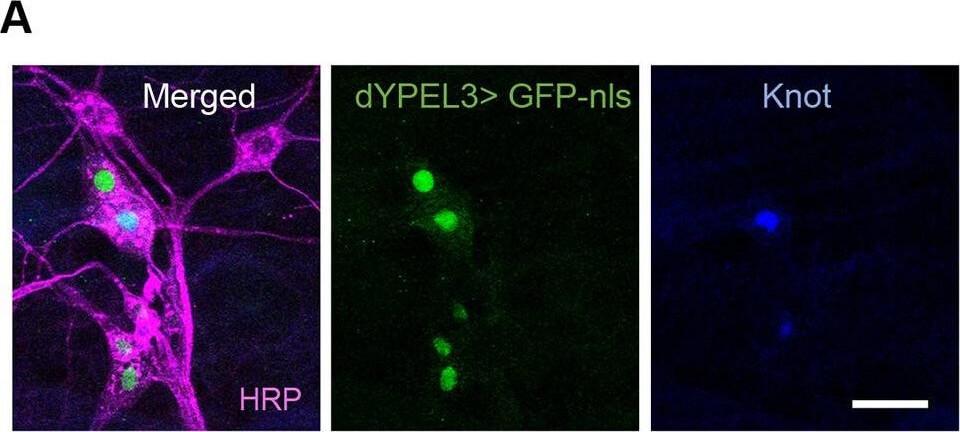
dYPEL3 frameshift mutations reduce nociceptive behavior. (A) Nociceptive/class IV da neurons are positive for dYPEL3. A nuclear GFP (GFP-nls, green) was expressed under dYPEL3-GAL4 following immunostaining with anti-Knot antibody (blue). Anti-HRP antibody was used to label all PNS neurons (magenta). Scale bar: 10 µm. (B) The AITC-induced nociceptive behavior was measured in a wild-type control (wt) and dYPEL3 frameshift mutants (dYPEL3T1-8 and dYPEL3T1-6). The number of larvae that exhibited complete rolling behavior was scored and expressed as a percentage (n=252 for each genotype). The Chi-squared test was performed between the groups. NS, non-significant, ****P<0.0001. Figure provided by CiteAb. Source: Dis Model Mech, PMID: 32461240.
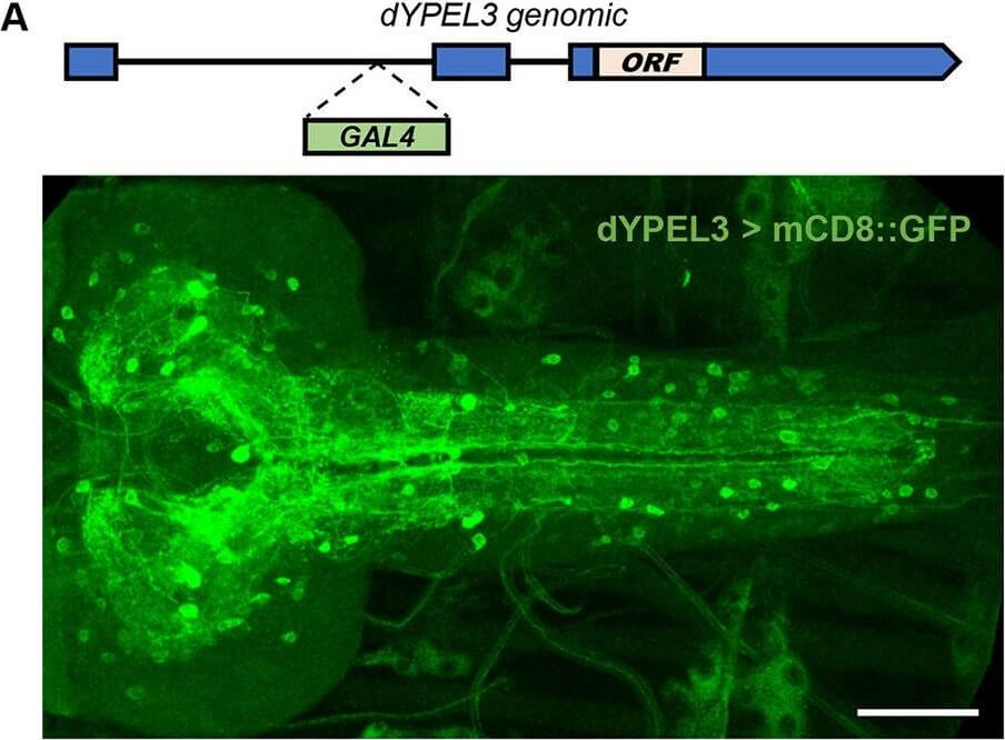
dYPEL3 is a neuronal gene. (A) The expression pattern of dYPEL3 in the CNS. The
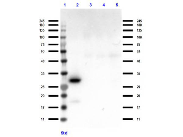
Western Blot of Ready-to-Use Rabbit Anti-RFP Antibody. Lane 1: Opal Prestain Molecular Weight (p/n MB-210-0500). Lane 2: RFP (p/n 000-001-379). Lane 3: Human IgG (p/n 009-0102). Lane 4: Goat IgG (p/n 005-0102). Lane 5: Mouse IgG (p/n 010-0102). Primary Antibody: RTU-RFP at 1µL/mL overnight at 4C. Secondary Antibody: Goat anti-Rabbit HRP (p/n 611-103-122) at 1:70,000 for 30min at RT. Expect 27kDa seen in lane 2.
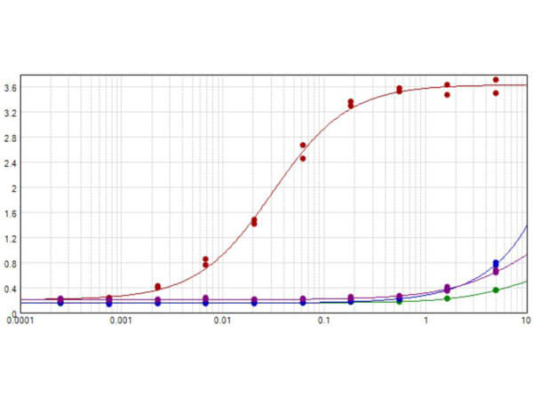
ELISA results of purified Ready-To-Use Rabbit Anti-RFP Antibody Min X Hu Ms and Rt Serum Proteins tested against RFP (Red), Mouse IgG (Blue), Human IgG (Green), and Rat IgG (Purple). Each well was coated in 1.0 µg of antigen. The starting dilution of antibody was 5 µg/mL and the X-axis represents the Log10 of a 3-fold dilution. This titration is a 4-parametre curve fit where the IC50 is defined as the titer of the antibody. The curve representing the RFP as the antigen is shown in red while Mouse IgG, Human IgG, and Rat IgG antigens are shown in blue, green, and purple. Assay performed using Buffer (p/n MB-060-1000), Substrate (p/n TMB-8000), and Conjugate (Goat Anti-Rabbit IgG Antibody HRP at 1:8,500).
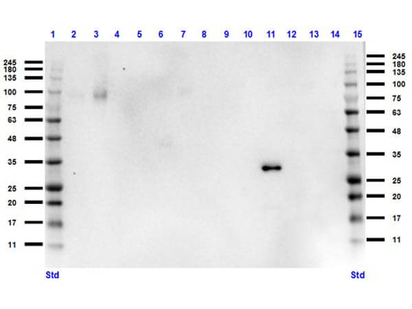
Western Blot of Ready-to-Use Rabbit Anti-RFP Antibody. Lane 1: Opal Prestain Molecular Weight (p/n MB-210-0500). Lane 2: HeLa (p/n W09-000-364). Lane 3: HEK293 (p/n W09-000-365). Lane 4: Cho/K1 (p/n W07-000-359). Lane 5: MDA-MB-231 (p/n W09-001-GK6). Lane 6: A431 (p/n W09-000-361). Lane 7: Jurkat (p/n W09-001-370). Lane 8: NIH/3T3 (p/n W10-000-358). Lane 9: E. coli HCP (p/n 000-001-J08). Lane 10: FLAG (p/n W00-001-383). Lane 11: RFP (p/n 000-001-379). Lane 12: GFP (p/n 000-001-215). Lane 13: GST (p/n 000-001-200). Lane 14: MBP (p/n 000-001-385). Lane 15: Opal Prestain. Primary Antibody: RTU-RFP at 1µL/mL overnight at 4C. Secondary Antibody: Goat anti-Rabbit HRP (p/n 611-103-122) at 1:70,000 for 30min at RT. Expect 27kDa seen in lane 11. Unspecific band in lane 3 caused by cross reactivity with secondary antibody.
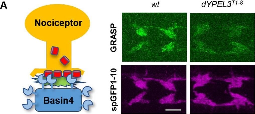
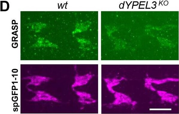
dYPEL3 frameshift mutations reduce the synaptic contact between nociceptors and Basin-4 neurons. (A) The syb-GRASP technique was used to report the synaptic contact between nociceptors and Basin-4. The spGFP1-10 (red cylinders) and spGFP11 (blue sectors) were expressed in nociceptors and Basin-4, respectively (left). The resulting GRASP signal was visualized by anti-GRASP antibody (green), and the spGFP1-10 that is expressed in nociceptor axon terminals was used as a normalization control (magenta) (right). Scale bar: 10µm. (B) The GRASP intensity from each neuropil was normalized by spGFP1-10 intensity and presented as means.e.m. (left), as well as in a violin plot to show distribution (right) (n=36 for wt, n=34 for dYPEL3T1-8). Mann-Whitney test. All statistical analysis was two-tailed. **P<0.01. Figure provided by CiteAb. Source: Dis Model Mech, PMID: 32461240.
* VAT and and shipping costs not included. Errors and price changes excepted