This antibody was purified from whole rabbit serum prepared by repeated immunizations with Myc epitope tag peptide, E-Q-K-L-I-S-E-E-D-L, conjugated to KLH using maleimide. The sequence corresponds to amino acids 410-419 of human c-Myc.
Conjugation:
Unconjugated
Alternative Names:
Rabbit anti-MYC Epitope Tag Antibody, Rabbit anti-c-myc, Glu-Gln-Lys-Leu-Ile-Ser-Glu-Glu-Asp-Leu
Clonality:
Polyclonal
Concentration:
1.0 mg/ml by UV absorbance at 280 nm
Buffer:
0.02 M Potassium Phosphate, 0.15 M Sodium Chloride, pH 7.2
Form:
Liquid (sterile filtered)
Antibody Type:
Primary Antibody
Application Dilute:
ELISA: 1:135,000, IHC: User Optimized, WB: 1:500 - 1:5,000
Application Notes:
Anti-Myc has utility to detect the fusion protein of the Myc epitope cloned along with the target gene. As such, anti-Myc/Myc can be used to identify fusion proteins containing the Myc epitope. The antibody recognizes the Myc tag fused either to the AMIN
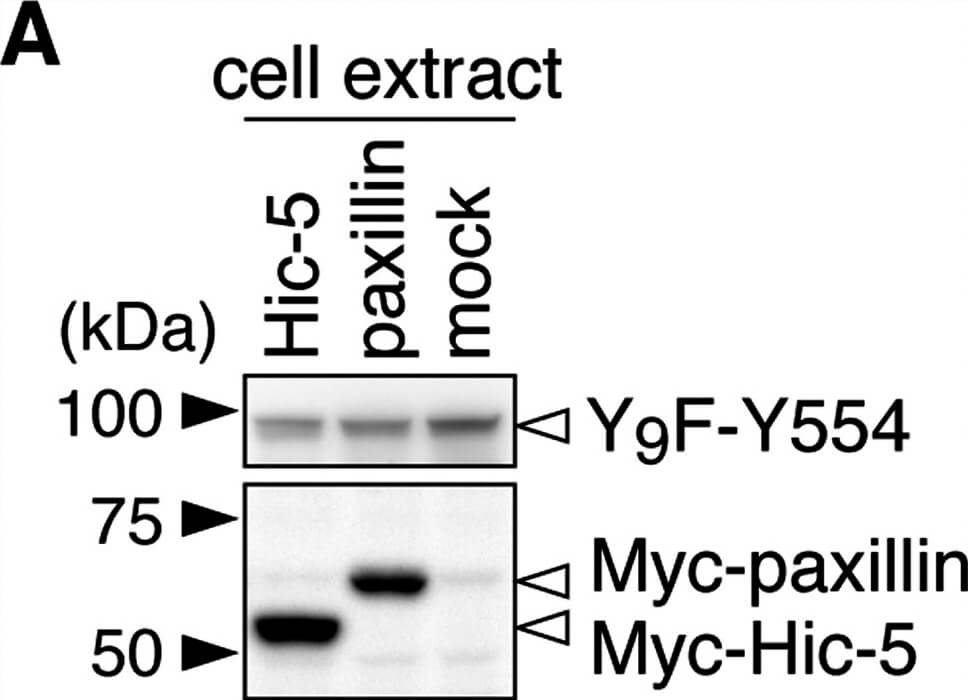
Git1 phosphorylation at Tyr-554 was enhanced by co-expression of paxillin.A, Western blotting of protein expression levels in HEK293T cells exogenously expressing the FLAG-tagged Git1-Y9F-Y554 mutant together with Myc-tagged paxillin, Myc-tagged Hic-5, or a control mock. B, Tyrosine phosphorylation of Y9F-Git1 proteins in anti-FLAG immunoprecipitates. The lower graph shows the densitometric analysis of the Western blotting data. Data are the mean S.E. (error bars, n = 3). *, P < 0.05 significantly different from Hic-5-transfected cells by ANOVA with Fishers PLSD post hoc tests. Figure provided by CiteAb. Source: PLoS One, PMID: 25742295.
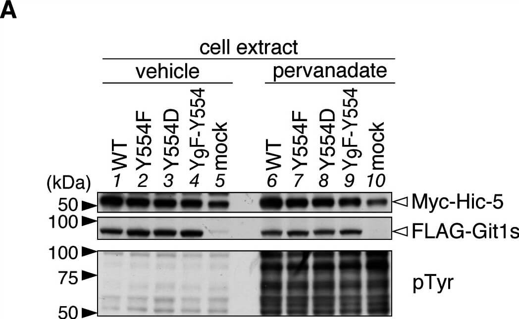
Git1 phosphorylation at Tyr-554 weakened its association with Hic-5.A, Western blotting of protein expression levels, and tyrosine phosphorylation of all proteins in HEK293T cells expressing FLAG-tagged Git1 proteins (Fig. 1A) together with Myc-tagged Hic-5. Cells were treated with 100 µM pervanadate or vehicle for 15 min, and then analyzed by Western blotting using anti-FLAG M2, anti-Myc 9E10, or anti-phosphotyrosine PY20. B, Co-immunoprecipitaion of Git1 mutants with Hic-5. The immunoprecipitates from cell extracts with anti-FLAG beads were analyzed by Western blotting with an anti-FLAG or anti-Myc antibody. To verify the tyrosine phosphorylation of FLAG-tagged Git1 proteins, the same membrane was reacted with anti-phosphotyrosine PY20. Ig, immunoglobulin. The lower part shows the densitometric analysis of the relative amount of Myc-Hic-5 to FLAG-Git1 in the immunoprecipitates. Data are the mean S.E. (error bars, n = 3). **, P < 0.01 significantly different from the wild-type with the same treatment, , P < 0.05 or , P < 0.01 significant difference between vehicle- and pervanadate-treated groups by ANOVA with Fishers PLSD post hoc tests. Figure provided by CiteAb. Source: PLoS One, PMID: 25742295.
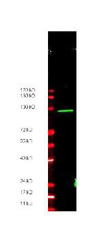
Anti-Myc epitope tag polyclonal antibody detects ~ 100 kDa CARBOXY terminal linked Myc-tagged recombinant protein present in ~35 µg of lysate by western blot. Carboxy terminal linked Myc recombinant protein was the gift of Zhongsheng You, Salk Institute, LaJolla, CA.
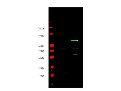
Anti-Myc epitope tag polyclonal antibody detects both AMINO and CARBOXY terminal linked Myc-tagged recombinant proteins by western blot. Polyclonal rabbit host anti-Myc epitope tag antibody was diluted to 1.0 µg/ml to detect either recombinant protein. 4-20% gradient gels were used to resolve the proteins by SDS-PAGE. The proteins were transferred to nitrocellulose using standard methods. After blocking, the membranes were probed with the primary antibody overnight at 4C followed by washes and reaction with a 1:10,000 dilution of IRDye 800 conjugated Gt-a-Rabbit IgG (H&L) MX10 (code 611-132-122) for 45 min at room temperature (Green, 800 nm channel). Pre-stained molecular weight markers are also shown (lane M, Red, 700 nm channel). LICORs Odyssey Infrared Imaging System was used to scan and process the image. Other detection systems will yield similar results.
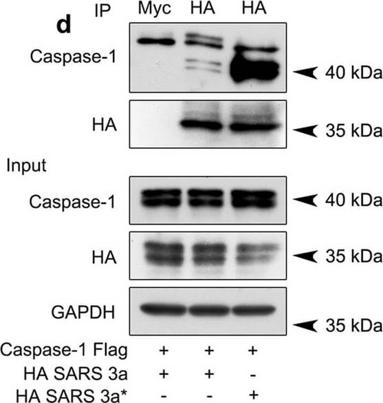
SARS 3a induces NLRP3 inflammasome activation by multiple mechanisms. A) Immunoblot analysis of the pro- and cleaved forms of caspase-1 and IL-1beta after reconstitution of inflammasome in HEK 293T cells transfected with SARS 3a with or without NEK7 shRNA. B) Immunoblot analysis of the pro- and cleaved forms of caspase-1 and IL-1beta after reconstitution of inflammasome and transfection with SARS 3a or SARS 3a C133A. C) Immunoblot analysis of the pro- and cleaved forms of caspase-1 and IL-1beta after co-transfection with caspase-1, IL-1beta, and SARS 3a or SARS 3a C133A. D) Immunoprecipitation analysis of interaction between SARS 3a or SARS 3a C133A and caspase-1. All western blot data are representative of two or three independent experiments Figure provided by CiteAb. Source: Cell Death Dis, PMID: 30185776.
* VAT and and shipping costs not included. Errors and price changes excepted