ATM Protein Kinase S1981 Antibody, Rabbit, Polyclonal
Biozol Catalog Number:
ROC-600-401-398
Supplier Catalog Number:
600-401-398
Alternative Catalog Number:
ROC-600-401-398
Manufacturer:
Rockland Immunochemicals
Host:
Rabbit
Category:
Antikörper
Application:
ELISA, IHC, WB
Species Reactivity:
Human, Mouse
Immunogen:
This antibody was affinity purified from whole rabbit serum prepared by repeated immunizations with a synthetic peptide S-L-A-F-E-E-G-S-Q-S-T-T-I-S-S corresponding to aa 1974-1988 of human ATM conjugated to KLH using maleimide.
0.02 M Potassium Phosphate, 0.15 M Sodium Chloride, pH 7.2
Form:
Liquid (sterile filtered)
Target:
Human
Antibody Type:
Primary Antibody
Application Dilute:
ELISA: 1:10,000 - 1:50,000, IHC: User Optimized, WB: 1:500- 1:2,000
Application Notes:
Affinity purified rabbit anti-ATM has been tested by ELISA, immunohistochemistry, western blotting and suitable for biological assays against both the native and recombinant forms of the protein.
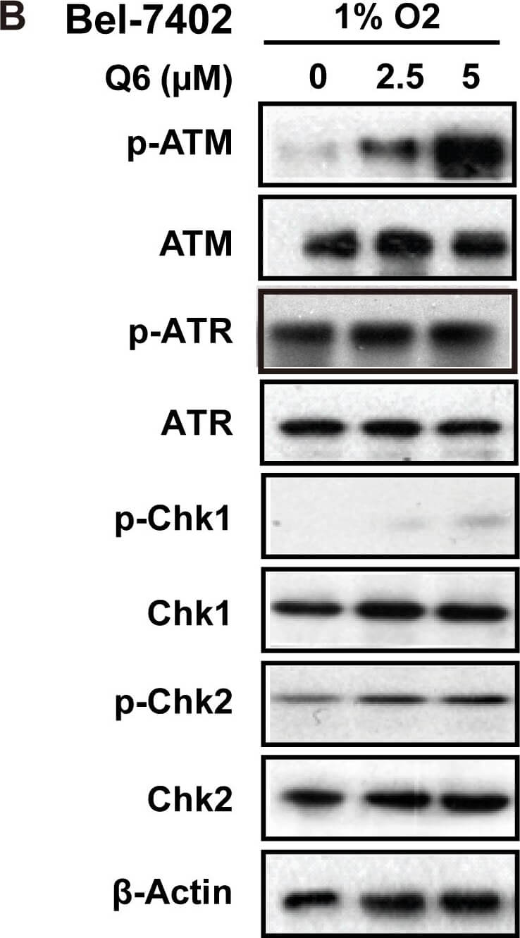
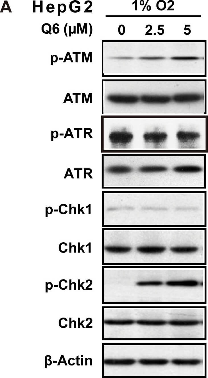
DNA DSBs induced by Q6 triggered ATM-Chk2 pathway in hypoxia.Western blot was carried out to explore the ATM/ATR signaling pathways in respond to DNA DSBs induced by Q6. A. HepG2 cells and B. Bel-7402 cells were treated with different concentration of Q6 (2.5 µM, 5 µM) under hypoxia (1% O2) condition. Protein levels were detected by Western blot analysis. beta-Actin was measured as the loading control. Data are representative of three independent experiments. Figure provided by CiteAb. Source: PLoS One, PMID: 26649750.
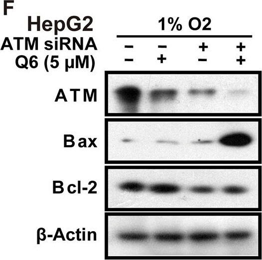
Q6 induced G2/M arrest and apoptosis is ATM/Chk2 dependent in hypoxia.A. HepG2 and Bel-7402 cells, treated with Q6 (5 µM) in the presence or absence of caffeine (2 mM) for 24 h under hypoxia (1% O2), were collected and prepared for cytometric analysis of cell cycle distribution. B & C. HepG2 cells treated with Q6 (10 µM) in the presence or absence of caffeine (2 mM) for 24 h under hypoxia (1% O2). Detection of apoptosis by flow cytometry (B) and caspase cascade by Western blot (C) were then performed. D & E. HepG2 cells treated with Q6 in the presence or absence of ATM specific inhibitor KU-60019 (3 µM) under hypoxia (1% O2), and subjected to sub-G1 analyses (D) and Western blot analyses (E), respectively. F. Western blot was used to assess the role of ATM during apoptosis induced by Q6 in hypoxia. HepG2 cells were treated with ATM RNAi or vector RNAi in the presence or absence of Q6 (5 µM) under hypoxia (1% O2) condition. Figure provided by CiteAb. Source: PLoS One, PMID: 26649750.
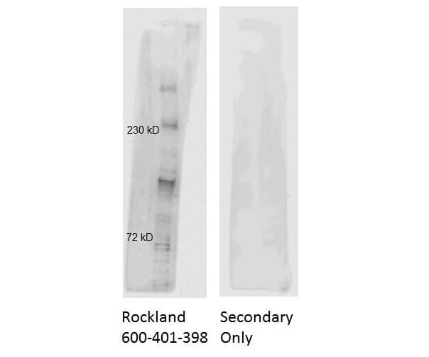
Western Blot of Rabbit Anti-ATM Antibody. HeLa Nuclear Lysates run on 4-8% gel and transferred for 1 hr at 100V to 0.45 µm nitrocellulose. Antibody Dilution Buffer: Buffer (p/n MB-070). Block: 5% Blotto for 1hr at 2-8C. Primary Antibody: Anti-ATM at 1:1000 overnight at 2-8C. Secondary Antibody: Goat Anti-Rabbit IgG HRP (p/n 611-103-122) at 1:20,000 for 1hr at 2-8C.
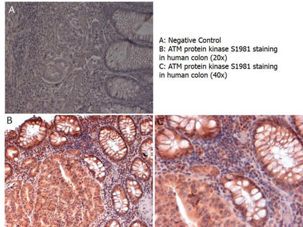
Immunohistochemistry with anti-ATM protein kinase S1981 antibody showing ATM protein kinase S1981 staining in nucleus and cytoplasm of human colon at 20x and 40x (B & C). Formalin fixed/paraffin embedded sections were subjected to heat induced epitope retrieval (HIER) at pH 6.2 and then incubated with rabbit anti-ATM protein kinase S1981 antibody at 4.0 µg/ml for 60 minutes. The reaction was developed using MACH 1 universal HRP polymer detection system and visualized with 33-diamino-benzidine substrate (DAB).
DNA DSBs induced by Q6 triggered ATM-Chk2 pathway in hypoxia.Western blot was carried out to explore the ATM/ATR signaling pathways in respond to DNA DSBs induced by Q6. A. HepG2 cells and B. Bel-7402 cells were treated with different concentration of Q6 (2.5 µM, 5 µM) under hypoxia (1% O2) condition. Protein levels were detected by Western blot analysis. beta-Actin was measured as the loading control. Data are representative of three independent experiments. Figure provided by CiteAb. Source: PLoS One, PMID: 26649750.
* VAT and and shipping costs not included. Errors and price changes excepted