Anti-Nestin affinity purified antibody was prepared from whole rabbit serum produced by repeated immunizations with a synthetic peptide corresponding to amino acids 1484-1500 of human Nestin protein.
This affinity purified antibody has been tested for use in ELISA, immunohistochemistry, and western blot. Specific conditions for reactivity should be optimized by the end user. Expect a band approximately 200-220 kDa in size corresponding to Nestin prot
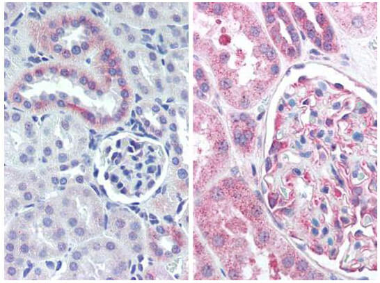
Immunohistochemistry with Anti-Nestin antibody. Tissue: mouse kidney (Left) and human kidney (Right). Fixation: formalin-fixed, paraffin-embedded tissue. Antigen retrieval: heat-induced. Primary antibody: 5 µg/ml. Staining: antibody as precipitated red signal with a hematoxylin purple nuclear counterstain.
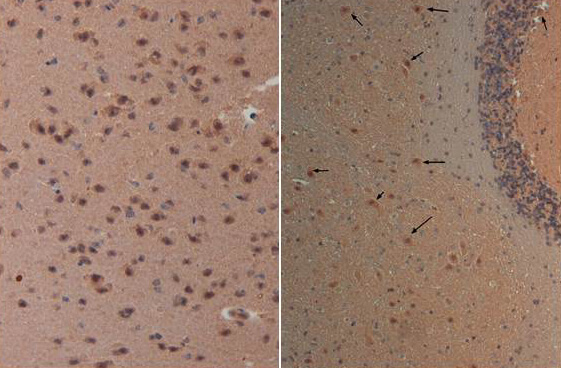
Immunohistochemistry with Anti-nestin antibody at 40X (left) and 20X (right) Tissue: Brain and Cerebellum (right) Fixation: FFPE buffered formalin 10% conc Antigen retrieval: Heat, Citrate pH 6.2. Pressure Cooker Primary antibody: 20ug/ml 1 hour room T Secondary antibody: Goat anti Rabbit Polymer HRP Prediluted by the manufacturer 30 min. room T Staining: antibody as precipitated red signal with a hematoxylin purple nuclear counterstain.
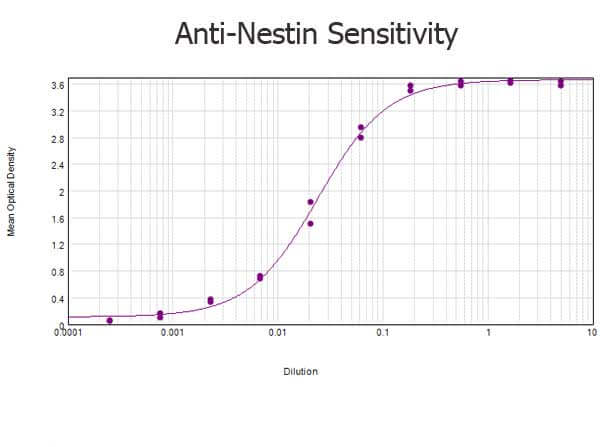
ELISA results of purified Rabbit anti-Nestin Antibody tested against BSA-conjugated peptide of immunizing peptide. Each well was coated in duplicate with 0.1µg of conjugate. The starting dilution of antibody was 5µg/ml and the X-axis represents the Log10 of a 3-fold dilution. This titration is a 4-parameter curve fit where the IC50 is defined as the titer of the antibody. Assay performed using 3% fish gel, Goat anti-Rabbit IgG Antibody Peroxidase Conjugated (Min X Bv Ch Gt GP Ham Hs Hu Ms Rt & Sh Serum Proteins) (p/n 611-103-122) and TMB ELISA Peroxidase Substrate (p/n TMBE-1000).
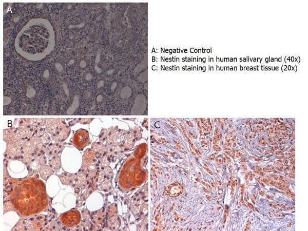
Immunohistochemistry with anti-nestin antibody showing nestin staining in cytoplasm of ductal epithelium of human salivary gland (B) and in nucleus and cytoplasm of human breast tissue (C). Formalin fixed/paraffin embedded sections were subjected to heat induced epitope retrieval (HIER) at pH 6.2 and then incubated with rabbit anti-nestin antibody at 4.0 µg/ml for 60 minutes. The reaction was developed using MACH 1 universal HRP polymer detection system and visualized with 33-diamino-benzidine substrate (DAB).
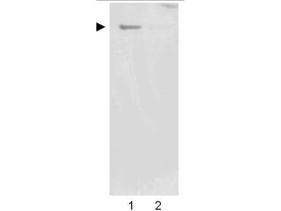
Western blot using Rocklands Affinity Purified anti-Nestin antibody shows detection of a band ~220 kDa corresponding to human Nestin (arrowhead). Undifferentiated HCN-1A human brain cortical neuron neuronal progenitor lysate (lane 1), or differentiated HCN-1A human brain cortical neuron neuronal progenitor lysate (lane 2) were separated by SDS-PAGE using 4-20% gradient gel. After transfer onto nitrocellulose, the membrane was blocked and then probed with the primary antibody diluted to 1:2,000 overnight at 4C. The membrane was then washed and reacted with a 1:10,000 dilution of peroxidase conjugated affinity purified Gt-a-Rabbit IgG [H&L] MX (611-1302) for 45 min at room temperature. Image was captured using film. Other detection systems will yield similar results. Image courtesy of Prof. F.H. Gage of the Salk Institute, San Diego, CA
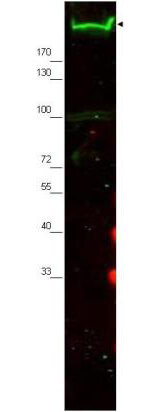
Western blot using Rocklands Affinity Purified anti-Nestin antibody shows detection of a band ~220 kDa corresponding to mouse Nestin (arrowhead). Approximately 30 g of MEF whole cell lysate was separated by SDS-PAGE using a 4-20% gradient gel. After transfer onto nitrocellulose, the membrane was blocked and then probed with the primary antibody diluted to 1:2,000 overnight at 4C. The membrane was then washed and reacted with a 1:10,000 dilution of IRDye(TM)800 conjugated Gt-a-Rabbit IgG [H&L] MX (611-132-122) for 45 min at room temperature. IRDye800 fluorescence image was captured using the Odyssey Infrared Imaging System developed by LI-COR. IRDye is a trademark of LI-COR, Inc. Other detection systems will yield similar results.
* VAT and and shipping costs not included. Errors and price changes excepted