PPAR alpha Antibody was prepared from whole rabbit serum produced by repeated immunizations with a synthetic peptide corresponding to a N-Terminal region near amino acids 1-25 of mouse PPAR alpha.
Conjugation:
Unconjugated
Alternative Names:
rabbit anti-Ppar alpha antibody, Pparalpha, Peroxisome proliferator-activated receptor alpha, PPAR-alpha, Nuclear receptor subfamily 1 group C member 1, Ppar-a, Nr1c1, Ppar
Anti-PPAR alpha Antibody has been tested in ELISA, Western Blot, Immunohistochemistry, and Immunofluorescence. Expect a single band approximately 52 kDa in size corresponding to PPAR alpha by western blot in the appropriate tissue or cell lysate. A 1:200
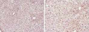
Immunohistochemistry (Formalin/PFA-fixed paraffin-embedded sections) showing Rocklands PPAR alpha antibody stainingof PPAR alphaprotein in mouse liver tissue section (Formalin/PFA-fixed paraffin-embedded sections). Tissue underwent formaldehyde fixation before enzymatic antigen retrieval with 0.05% protease in PBS for5 minutes. Sample was then blockedwith5% serum for20 minutes at 20C. The primary antibody was diluted 1:50 and incubated with sample in Tris plus 5% normal goat serum for 1 hour at 20C. ABiotin conjugatedgoat polyclonal to rabbit IgG was used at dilution at1:500as secondary antibody. Images show nuclear staining in hepatocytes (perfusion-fixed mouse, 10 and 40x microscope magnification).
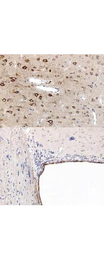
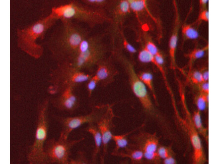
Immunohistochemistry using Rocklands anti-PPAR antibody, showing staining of PPAR alpha in rat brain sections, highlighting cytoplasmic staining in ependymal cells and neurons in frontal cortex. Bottom image shows subventricular zone (svz) of lateral ventrical (exit point of progenitor olfactory neurones), top image shows frontal cortex in the same section. Cytoplasmic staining is also observed in the corpus callosum (bottom image) and in dendritic fields of the cortex. Formalin/PFA-fixed paraffin-embedded sections of rat brain tissue were incubated with the primary antibody at 1:200 for 1 hour. Antigen retrieval was performed by heat induction in citrate buffer pH 6.0.
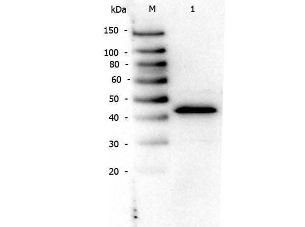
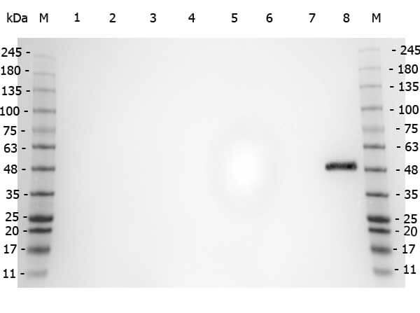
Western Blot of Rabbit anti-PPAR Alpha (N-terminal Specific) antibody. Lane M: Prestained Molecular Weight Markers. Lane 1: NIH/3T3 (p/n W10-000-358). Load: 10 µg per lane. Primary antibody: PPAR Alpha (N-terminal specific) antibody at 1:1,000 for overnight at 4C. Secondary antibody: Peroxidase rabbit secondary antibody (p/n 611-103-122) at 1:40,000 for 30 min at RT. Block: Blocking Buffer for Fluorescent Western Blotting (p/n MB-070) at RT for 30 min. Predicted/Observed size: ~50 kDa for PPAR Alpha.
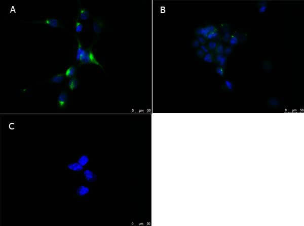
Immunofluorescence microscopy of Rabbit Anti-PPAR alpha (N-terminal spec
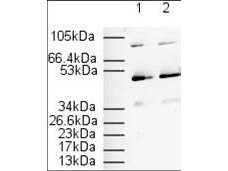
Western Blot of Rabbit anti-PPAR Alpha (N-terminal specific) antibody. Marker: Opal Pre-stained ladder (p/n MB-210-0500). Lane 1: HEK293 lysate (p/n W09-000-365). Lane 2: HeLa Lysate (p/n W09-000-364). Lane 3: MCF-7 Lysate (p/n W09-000-360). Lane 4: Jurkat Lysate (p/n W09-000-370). Lane 5: A431 Lysate (p/n W09-000-361). Lane 6: LNCaP Lysate (p/n W09-001-GJ9). Lane 7: A-172 Lysate (p/n W09-001-GL5). Lane 8: NIH/3T3 Lysate (p/n W10-000-358). Load: 35 µg per lane. Primary antibody: PPAR Alpha (N-terminal specific) antibody at 1ug/mL overnight at 4C. Secondary antibody: Peroxidase rabbit secondary antibody (p/n 611-103-122) at 1:30,000 for 60 min at RT. Blocking Buffer: 1% Casein-TTBS (p/n MB-082) for 30 min at RT. Predicted/Observed size: 52 kDa for PPAR Alpha.
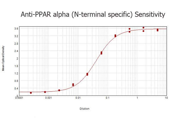
ELISA results of purified Rabbit anti-PPAR Alpha (N-terminal specific) Antibody tested against BSA-conjugated peptide of immunizing peptide. Each well was coated in duplicate with 0.1µg of conjugate. The starting dilution of antibody was 5µg/ml and the X-axis represents the Log10 of a 3-fold dilution. This titration is a 4-parameter curve fit where the IC50 is defined as the titer of the antibody. Assay performed using 3% fish gel, Goat anti-Rabbit IgG Antibody Peroxidase Conjugated (Min X Bv Ch Gt GP Ham Hs Hu Ms Rt & Sh Serum Proteins) (p/n 611-103-122) and TMB ELISA Peroxidase Substrate (p/n TMBE-1000).
Immunofluorescence Microscopy of Rabbit anti-PPAR alpha antibody. Tissue: HepG2 cells. Fixation: 4% formaldehyde fixed (10 min). Antigen retrieval: not required. Primary antibody: PPAR alpha antibody at 1 µg/mL overnight at 4C. Secondary antibody: Alexa Fluor 488 goat anti-rabbit IgG (H+L) (green) used at a 1:1000, Alexa Fluor 594 WGA was used to label plasma membranes (red) at a 1:200 dilution for 1h for 45 min at RT. Localization: PPAR alpha is nuclear and occasionally cytoplasmic. Staining: PPAR alpha as green fluorescent signal with DAPI (blue) nuclear counterstain.
* VAT and and shipping costs not included. Errors and price changes excepted