This affinity purified antibody was prepared from whole rabbit serum produced by repeated immunizations with a synthetic peptide corresponding to an internal region near amino acids 115-140 of Human Cyclin B1 protein.
0.02 M Potassium Phosphate, 0.15 M Sodium Chloride, pH 7.2
Form:
Liquid (sterile filtered)
Target:
Human
Antibody Type:
Primary Antibody
Application Dilute:
ELISA: 1:50,000, IP: 1:100, WB: 1:100 - 1:1,000
Application Notes:
This affinity purified antibody has been tested for use in ELISA and western blot. Specific conditions for reactivity should be optimized by the end user. Expect a band ~ 48 kDa in size corresponding to Cyclin B1 by western blotting in the appropriate ce
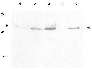
Western blot using Rocklands Affinity Purified anti-Cyclin B1 pS126 antibody shows detection of a band ~48 kDa corresponding to phosphorylated human Cyclin B1 (arrowheads) in various whole cell lysates. Lane 1: Hela (cervical carcinoma) [p/n W09-000-364], Lane 2: H23 (lung carcinoma), Lane 3: Hep3b (Hepatocarcinoma), Lane 4: T98G (Glioblastoma), and Lane 5: Daudi (B cell lymphoblast) [W09-001-MQ2]. Each lane contains approximately 50 µg of lysates, separated by 12% SDS-PAGE using a 5% stack run at 100 volts until the dye front cleared the bottom of the gel. Transfer occurred overnight at 4C at 15 mAmps. The membrane was blocked with 5% non-fat dry milk in TTBS (p/n B501-0500) for 1 h at room temperature followed by addition of a 1:100 dilution of the antibody allowed to react for 2h at room temperature. After washes with TTBS a 1:5,000 dilution of HRP conjugated Gt-a-Rabbit IgG [H&L] MX (p/n 611-103-122) was added for 1 h at room temperature. After additional washes the membrane was incubated with ECL mix 1:1 for ~3 min. Excess detection solution was drained off and the membrane was exposed to Kodak film X-omat blue XB-1 for about 20 sec. Other detection systems will yield similar results. Personnel Communication, Luca Cote, Temple U.
* VAT and and shipping costs not included. Errors and price changes excepted