AURORA KINASE B phospho T232 Antibody, Rabbit, Polyclonal
Biozol Catalog Number:
ROC-600-401-677S
Supplier Catalog Number:
600-401-677S
Alternative Catalog Number:
ROC-600-401-677S
Manufacturer:
Rockland Immunochemicals
Host:
Rabbit
Category:
Antikörper
Application:
ELISA, IHC, WB
Species Reactivity:
Human, Monkey
Immunogen:
This affinity purified antibody was prepared from whole rabbit serum produced by repeated immunizations with a synthetic peptide corresponding to an internal region surrounding T232 of Human Aurora Kinase B protein.
Conjugation:
Unconjugated
Alternative Names:
rabbit anti-Aurora B pT232 Antibody, AIK2 antibody, AIM1 antibody, ARK2 antibody, AurB antibody, AURKB antibody, Aurora 1 antibody, Aurora and Ipl1 like midbody associated protein 1 antibody, Aurora Kinase B, IPL1-related kinase 2, Aurora-related kinase 2, STK-1, Aurora-B, Ipl1-like midbody-associated protein 1, AIM-1, Aurora/IPL1-related kinase 2
0.02 M Potassium Phosphate, 0.15 M Sodium Chloride, pH 7.2
Form:
Liquid (sterile filtered)
Target:
Human
Antibody Type:
Primary Antibody
Application Dilute:
ELISA: 1:10,000 - 1:50,000, IF Microscopy: User Optimized, WB: 1:250 - 1:2,000
Application Notes:
Anti-Aurora B pT232 Antibody is affinity-purified antibody has been tested for use in ELISA, immunohistochemistry, and by western blot. Expect a band approximately 39 kDa in size corresponding to Aurora Kinase B by western blotting in the appropriate cel
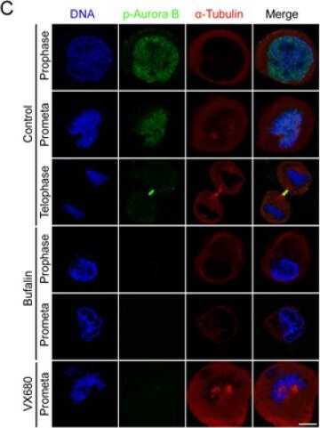
Bufalin prevents Aurora A recruitment to mitotic centrosomes and Aurora B recruitment to unattached kinetochores(A) HeLa cells were synchronized by a single thymidine treatment, released in the presence or absence of bufalin (100 nM) for 9 h, and stained for phospho-Aurora A (Green), alpha-tubulin (Red) and DNA (Blue). The scale bar represents 10 µm. (B) The phospho-Aurora A (Thr288) staining signals in (A) were normalized to the intensity in a same-size cytoplasmic region for at least five prometaphase cells per condition from three different experiments. ***p < 0.001, versus control prometaphase. Error bar represents SEM. (C) Thymidine-synchronized HeLa cells were treated with or without bufalin (100 nM) for 9 h and then stained for phospho-Aurora B (Green), alpha-tubulin (Red) and DNA (Blue). The scale bar represents 10 µm. (D) For quantification of the intensity of phospho-Aurora B (Thr232) in (C), m
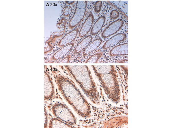
Immunohistochemistry of Rabbit Anti-AuroraB pT232 Antibody. Tissue: human intestine pH9 (A) at 20x and 40x. Fixation: formalin fixed paraffin embedded. Antigen retrieval: not required. Primary antibody: AuroraB pT232 antibody at 10 µg/mL for 1 h at RT. Secondary antibody: Peroxidase rabbit secondary antibody at 1:10,000 for 45 min at RT. Localization: AuroraB pT232 is cytoplasmic. Staining: AuroraB pT232 as precipitated brown signal with hematoxylin purple nuclear counterstain.
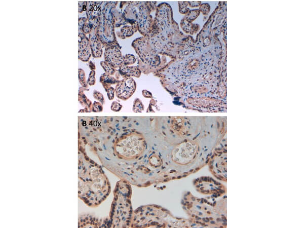
Immunohistochemistry of Rabbit Anti-AuroraB pT232 Antibody. Tissue: human placenta pH9 (A) at 20x and 40x. Fixation: formalin fixed paraffin embedded. Antigen retrieval: not required. Primary antibody: AuroraB pT232 antibody at 10 µg/mL for 1 h at RT. Secondary antibody: Peroxidase rabbit secondary antibody at 1:10,000 for 45 min at RT. Localization: AuroraB pT232 is cytoplasmic. Staining: AuroraB pT232 as precipitated brown signal with hematoxylin purple nuclear counterstain.
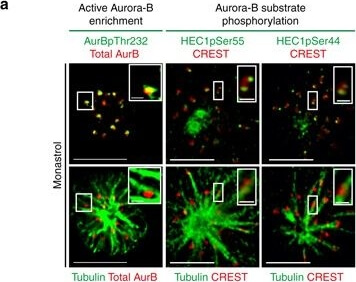
High Aurora-B activity promotes KT attachment to MT-walls. a Representative images show high Aurora-B activity on lateral kinetochores. Monastrol treated cells were immunostained with antibodies against Tubulin, Aurora-BpThr232 and total Aurora-B (AurB) (left panel) or CREST antisera and antibodies against Tubulin and either HEC1pSer55 (middle panel) or HEC1pSer44 (right panel). Cropped images show lateral-kinetochores. Scale: 5µm in uncropped images, 1µm in cropped images. b Graphs show higher average signal intensities of HEC1pSer55 (left) and HEC1pSer44 (right) in lateral compared to end-on kinetochores as assessed from at least nine randomly chosen kinetochores from cells in a. CREST signal intensities are used as internal controls. c Experimental regime: Cells transfected with plasmid vectors encoding Mis12-INCENP-GFP were exposed to Monastrol and MG132 with either ZM447439 or DMSO (solvent control), prior to immunostaining. d Images of cells expressing Mis12-INCENP-GFP treated as in c and immunostained with antibodies against Tubulin, SKAP and GFP. White arrowheads in cropped images show 'Lateral kinetochore lacking SKAP (upper panel) and 'End-on kinetochore enriched with SKAP (lower panel). Scale: 5µm in uncropped and 2µm in cropped images. Boxed areas in a and d correspond to cropped images. e Graph shows percentage of lateral, end-on and detached kinetochores in Mis12-INCENP-GFP expressing cells treated as in c. Each circle represents value from one cell. Black horizontal bar marks average values from three independent experimental repeats. '* indicates statistically significant difference on the basis of P-values obtained using unpaired Students t-test Figure provided by CiteAb. Source: Nat Commun, PMID: 28751710.
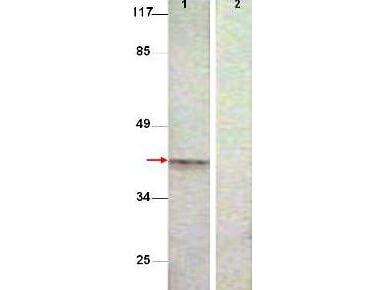
Western Blot shows detection of Aurora B protein at 39 kDa (predicted band size). All lanes : Aurora B (phospho T232) antibody diluted 1:500. Lane 1 : Extract from COS7 cells treated with Nocodazole (1ug/ml, 16 hrs). Lane 2 : Extract from COS7 cells treated with Nocodazole (1ug/ml, 16 hrs) and with the phosphopeptide immunogen.
* VAT and and shipping costs not included. Errors and price changes excepted