This affinity-purified antibody was prepared from whole rabbit serum produced by repeated immunizations with a synthetic peptide corresponding to an internal region near amino acids 85-115 of Human EGR-1.
Conjugation:
Unconjugated
Alternative Names:
rabbit anti-EGR-1 Antibody, EGR1, EGR 1, AT225 antibody, Early growth response 1 antibody, KROX24 antibody, Nerve growth factor-induced protein A antibody, NGFI-A, Transcription factor ETR103, Transcription factor Zif268, ZNF225, Zinc finger protein 225, Zinc finger protein Krox-24
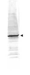
This affinity purified antibody has been tested for use in ELISA, immunohistochemistry and western blot. Specific conditions for reactivity should be optimized by the end user. Expect a band at ~58 kDa in size corresponding to EGR-1 by western blotting i
Western blot using Rocklands Affinity Purified anti-EGR-1 antibody shows detection of a predominant band at ~58 kDa corresponding to EGR-1 present in mouse embryonic fibroblast whole cell lysate (p/n W10-001-371) (arrowhead). Approximately 35 µg of lysate was separated by 4-20% SDS-PAGE and transferred onto nitrocellulose. After blocking the membrane was probed with the primary antibody diluted to 1:1,500. Reaction occurred 2h at room temperature followed by washes and reaction with a 1:10,000 dilution of IRDye(TM)800 conjugated Gt-a-Rabbit IgG [H&L] MX (p/n 611-132-122) for 45 min at room temperature. IRDye(TM)800 fluorescence image was captured using the Odyssey Infrared Imaging System developed by LI-COR. IRDye is a trademark of LI-COR, Inc. Other detection systems will yield similar results.
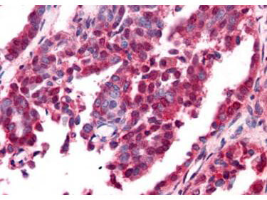
Rocklands Affinity Purified anti-EGR-1 antibody was used at a 10 ug/ml to detect nuclear and cytoplasmic signal with low background staining in a variety of tissues including multi-human, multi-brain and multi-cancer slides. Within the multi-tumor block, the antibody showed variable levels of nuclear and cytoplasmic staining between individual tumors, with some tumors showing moderate staining. This image shows EGR-1 staining of human ovarian carcinoma. Tissue was formalin-fixed and paraffin embedded. Personal Communication, Tina Roush, LifeSpanBiosciences, Seattle, WA.
* VAT and and shipping costs not included. Errors and price changes excepted